Keratoacanthoma,light micrograph
Bildnummer 11677191
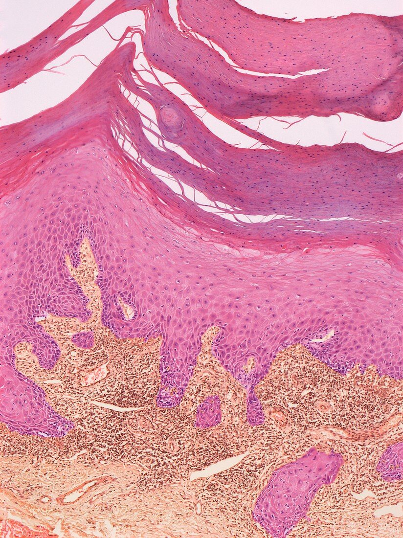
| Keratoacanthoma. Light micrograph of a section through a keratoacanthoma skin lesion. The surface layers are across top,including the hard keratin layer. The lesion consists of a localised proliferation of squamous cells that forms a cratered nodule. The nodule grows over several weeks before gradually disappearing. However,the unsightly nodule is often surgically removed. The cause of keratoacanthoma is unknown,although exposure to sunlight appears to be a factor. Magnification: x60 when printed at 10 centimetres wide. Human tissue | |
| Lizenzart: | Lizenzpflichtig |
| Credit: | Science Photo Library / Gschmeissner, Steve |
| Bildgröße: | 3426 px × 4572 px |
| Modell-Rechte: | nicht erforderlich |
| Eigentums-Rechte: | nicht erforderlich |
| Restrictions: | - |
Preise für dieses Bild ab 15 €
Universitäten & Organisationen
(Informationsmaterial Digital, Informationsmaterial Print, Lehrmaterial Digital etc.)
ab 15 €
Redaktionell
(Bücher, Bücher: Sach- und Fachliteratur, Digitale Medien (redaktionell) etc.)
ab 30 €
Werbung
(Anzeigen, Aussenwerbung, Digitale Medien, Fernsehwerbung, Karten, Werbemittel, Zeitschriften etc.)
ab 55 €
Handelsprodukte
(bedruckte Textilie, Kalender, Postkarte, Grußkarte, Verpackung etc.)
ab 75 €
Pauschalpreise
Rechtepakete für die unbeschränkte Bildnutzung in Print oder Online
ab 495 €
Keywords
- abnormal,
- dermal,
- Dermatologie,
- dermatologisch,
- epidermal,
- Epidermis,
- Gesundheitswesen,
- gutartig,
- Haut,
- Histopathologie,
- histopathologisch,
- Keratin,
- Keratoakanthom,
- Kondition,
- Krankheit,
- Lichtmikroskop,
- lichtmikroskopische Aufnahme,
- Masse,
- Medizin,
- medizinisch,
- Mensch,
- menschlicher Körper,
- menschliches Gewebe,
- nicht krebserregend,
- Niemand,
- Oberfläche,
- Sektion,
- sektioniert,
- Störung,
- ungesund,
- Wachstum,
- weißes Blutkörperchen,
- Wunde