White oak leaf,light micrograph
Bildnummer 11675913
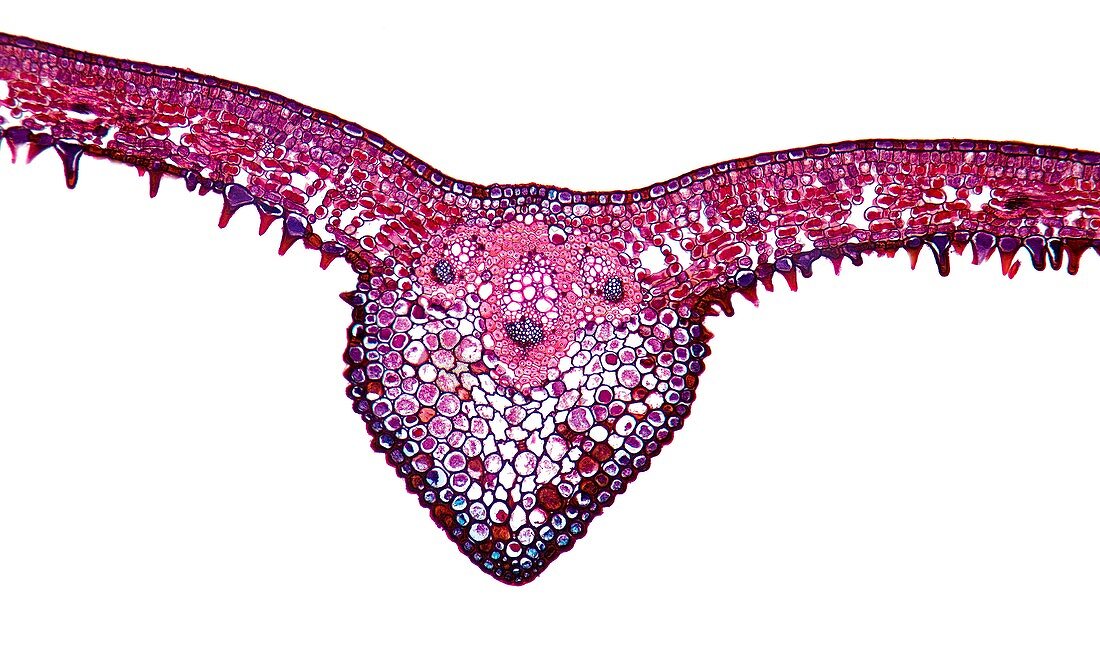
| White oak leaf. Light micrograph of a section through the midrib of a leaf from a white oak (Quercus alba) tree. This is a typical mesophyte leaf micrograph upper and lower epidermal layers (red-blue),a cuticle and typical stomata,found only on the lower surface. Under the upper epidermis are two layers of palisade mesophyll (red-violet) with chloroplasts. Beneath this is spongy mesophyll (red) with chloroplasts and large inter-cellular spaces. In the middle of the lamina is the midrib,carrying three large veins surrounded by parenchyma (violet),which are themselves surrounded by fibre/sclerenchyma (pink). Inside the top of the veins is the xylem (pink) with large tracheid cells. The lower veins are the phloem,with sieve tubes (small dark blue | |
| Lizenzart: | Lizenzpflichtig |
| Credit: | Science Photo Library / Wheeler, Dr. Keith |
| Bildgröße: | 5747 px × 3378 px |
| Modell-Rechte: | nicht erforderlich |
| Eigentums-Rechte: | nicht erforderlich |
| Restrictions: | - |
Preise für dieses Bild ab 15 €
Universitäten & Organisationen
(Informationsmaterial Digital, Informationsmaterial Print, Lehrmaterial Digital etc.)
ab 15 €
Redaktionell
(Bücher, Bücher: Sach- und Fachliteratur, Digitale Medien (redaktionell) etc.)
ab 30 €
Werbung
(Anzeigen, Aussenwerbung, Digitale Medien, Fernsehwerbung, Karten, Werbemittel, Zeitschriften etc.)
ab 55 €
Handelsprodukte
(bedruckte Textilie, Kalender, Postkarte, Grußkarte, Verpackung etc.)
ab 75 €
Pauschalpreise
Rechtepakete für die unbeschränkte Bildnutzung in Print oder Online
ab 495 €
Keywords
- Anatomie,
- anatomisch,
- Angiosperme,
- Angiospermen,
- Biologie,
- biologisch,
- Blatt,
- Botanik,
- botanisch,
- Chloroplasten,
- epidermal,
- Epidermis,
- Flora,
- Gefäß,
- Gewebe,
- Histologie,
- histologisch,
- Kutikula,
- lichtmikroskopische Aufnahme,
- Mesophyt,
- Mikroskop,
- Mittelrippe,
- Natur,
- niedriger,
- Pflanze,
- Pflanzen,
- Phloem,
- Schicht,
- Schichten,
- Sektion,
- sektioniert,
- Stomata,
- Struktur,
- Tierwelt,
- Vene,
- Venen,
- weißer Hintergrund,
- Xylem,
- Zelle,
- Zellen