Cardiac muscle,fluorescence micrograph
Bildnummer 11675585
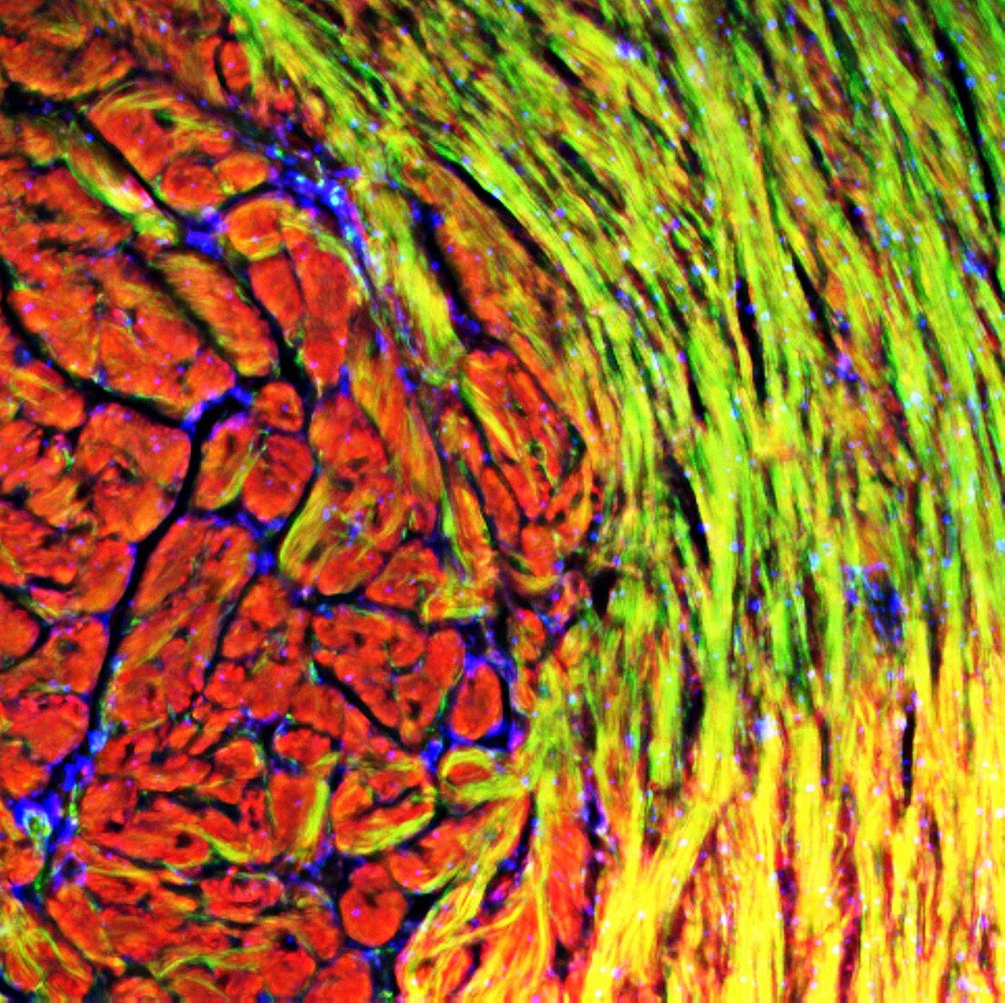
| Cardiac muscle. Fluorescence deconvolution micrograph of a section through cardiac (heart) muscle tissue. The muscle tissue is seen in both transverse (left) and longitudinal (right) sections. Cellular proteins are highlighted with fluorescent markers: g-actin (red),f-actin (green),and smooth muscle actin (blue). Magnification: x200 when printed at 10 centimetres across | |
| Lizenzart: | Lizenzpflichtig |
| Credit: | Science Photo Library / R. BICK, B. POINDEXTER, UT MEDICAL SCHOOL |
| Bildgröße: | 2982 px × 2976 px |
| Modell-Rechte: | nicht erforderlich |
| Eigentums-Rechte: | nicht erforderlich |
| Restrictions: | - |
Preise für dieses Bild ab 15 €
Universitäten & Organisationen
(Informationsmaterial Digital, Informationsmaterial Print, Lehrmaterial Digital etc.)
ab 15 €
Redaktionell
(Bücher, Bücher: Sach- und Fachliteratur, Digitale Medien (redaktionell) etc.)
ab 30 €
Werbung
(Anzeigen, Aussenwerbung, Digitale Medien, Fernsehwerbung, Karten, Werbemittel, Zeitschriften etc.)
ab 55 €
Handelsprodukte
(bedruckte Textilie, Kalender, Postkarte, Grußkarte, Verpackung etc.)
ab 75 €
Pauschalpreise
Rechtepakete für die unbeschränkte Bildnutzung in Print oder Online
ab 495 €
Keywords
- Anatomie,
- anatomisch,
- Biologie,
- biologisch,
- diagonal,
- Entfaltung,
- F-Actin,
- Farbstoff,
- Farbstoffe,
- Fasern,
- Flecken,
- Fluoreszenz,
- fluoreszierend,
- g-Actin,
- gesund,
- Gewebe,
- globuläres Aktin,
- Herz,
- Histologie,
- histologisch,
- Lichtmikroskop,
- lichtmikroskopische Aufnahme,
- Marker,
- menschlicher Körper,
- normal,
- Proteine,
- Querschnitt,
- Sektion,
- sektioniert,
- Verfärbung,
- Zellbilogie,
- Zellen,
- zellular,
- zelluläres Protein,
- Zytologie,
- Zytologisch,
- Zytoskelett