Healthy knee,CT scan
Bildnummer 11659023
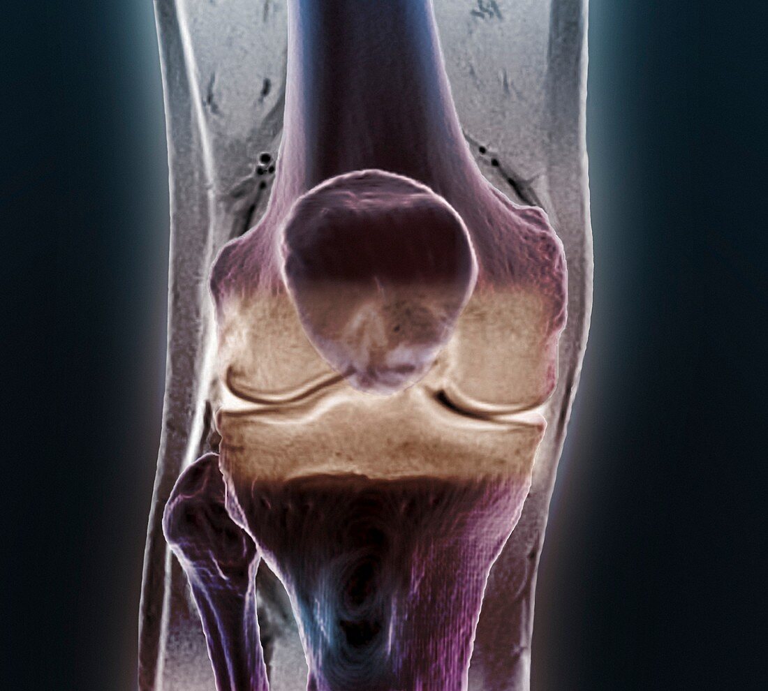
| Healthy knee. Coloured frontal computed tomography (CT) scan projected on to a magnetic resonance imaging (MRI) scan of the knee of a 30 year old. The knee joint is formed by the articulation of the femur (thigh bone,top) with tibia (shin bone,bottom centre). The smaller fibula (bottom left) is also seen. The patella (knee cap) is seen over the bottom of the femur | |
| Lizenzart: | Lizenzpflichtig |
| Credit: | Science Photo Library / Zephyr |
| Bildgröße: | 4412 px × 3969 px |
| Modell-Rechte: | nicht erforderlich |
| Eigentums-Rechte: | nicht erforderlich |
| Restrictions: | - |
Preise für dieses Bild ab 15 €
Universitäten & Organisationen
(Informationsmaterial Digital, Informationsmaterial Print, Lehrmaterial Digital etc.)
ab 15 €
Redaktionell
(Bücher, Bücher: Sach- und Fachliteratur, Digitale Medien (redaktionell) etc.)
ab 30 €
Werbung
(Anzeigen, Aussenwerbung, Digitale Medien, Fernsehwerbung, Karten, Werbemittel, Zeitschriften etc.)
ab 55 €
Handelsprodukte
(bedruckte Textilie, Kalender, Postkarte, Grußkarte, Verpackung etc.)
ab 75 €
Pauschalpreise
Rechtepakete für die unbeschränkte Bildnutzung in Print oder Online
ab 495 €
Keywords
- 30er Jahre,
- Anatomie,
- anatomisch,
- Bein,
- Biologie,
- biologisch,
- CT-Scan,
- dreißig,
- dreißiger Jahre,
- Erwachsene,
- farbig,
- Femur,
- Frontal,
- gefärbt,
- Gelenk,
- gesund,
- Joint,
- Knie,
- Knochen,
- Menschen,
- menschlicher Körper,
- MRT-Untersuchung,
- normal,
- Oberschenkelknochen,
- Person,
- Radiographie,
- Röntgen,
- Röntgengerät,
- Scanner,
- Schienbein,
- Vorderseite,
- Wadenbein,
- Zusammengesetzt