Chorionic villous sampling in pregnancy
Bildnummer 11656136
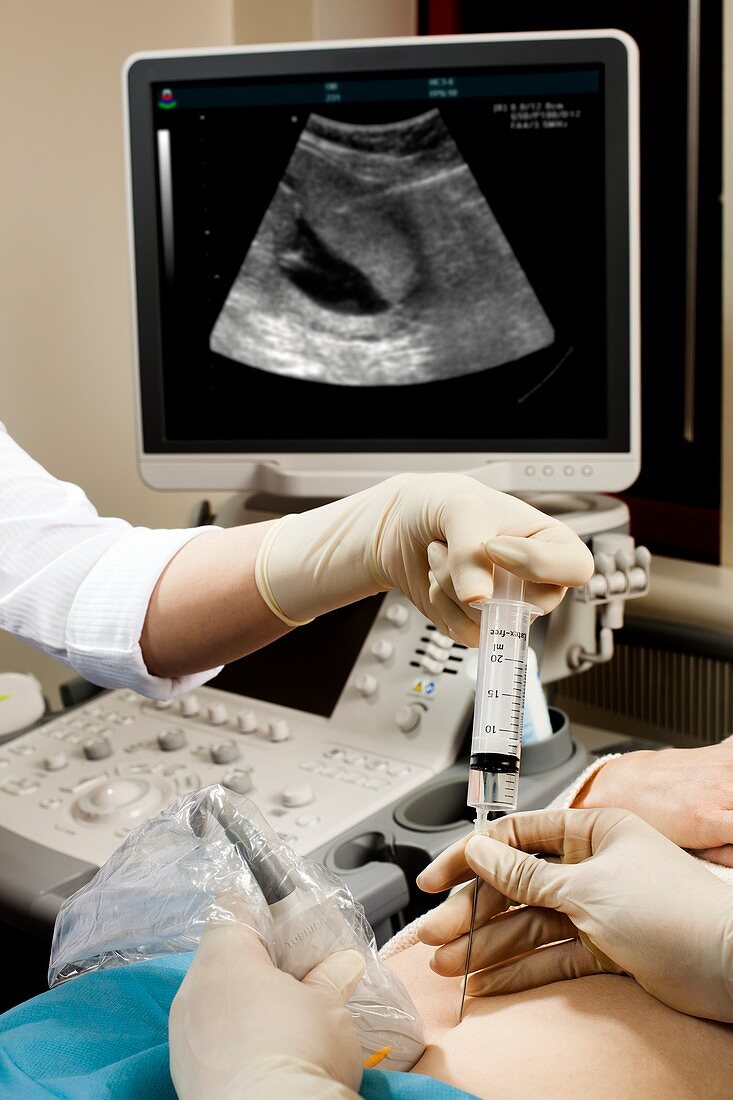
| MODEL RELEASED. Chorionic villous sampling in pregnancy. Medical staff carrying out chorionic villous sampling (CVS) on a pregnant woman. An ultrasound transducer has been used to guide the needle and syringe being used. The uterus,foetus and needle are seen on the ultrasound scan on the screen in the background. The foetal chorionic villi tissue,in the mother's placenta,contains foetal blood vessels and is a way of obtaining foetal blood. Chorionic villus sampling is usually conducted between 9 and 12 weeks of pregnancy to evaluate the chromosomal and DNA (deoxyribonucleic acid) status of the foetus. It is a way of diagnosing foetal abnormalities in early pregnancy | |
| Lizenzart: | Lizenzpflichtig |
| Credit: | Science Photo Library / SATURN STILLS |
| Bildgröße: | 3419 px × 5128 px |
| Modell-Rechte: | vorhanden |
| Eigentums-Rechte: | nicht erforderlich |
| Restrictions: | - |
Preise für dieses Bild ab 15 €
Universitäten & Organisationen
(Informationsmaterial Digital, Informationsmaterial Print, Lehrmaterial Digital etc.)
ab 15 €
Redaktionell
(Bücher, Bücher: Sach- und Fachliteratur, Digitale Medien (redaktionell) etc.)
ab 30 €
Werbung
(Anzeigen, Aussenwerbung, Digitale Medien, Fernsehwerbung, Karten, Werbemittel, Zeitschriften etc.)
ab 55 €
Handelsprodukte
(bedruckte Textilie, Kalender, Postkarte, Grußkarte, Verpackung etc.)
ab 75 €
Pauschalpreise
Rechtepakete für die unbeschränkte Bildnutzung in Print oder Online
ab 495 €
Keywords
- 2013,
- 21. Jahrhundert,
- Anzeige,
- Ausrüstung,
- Bildschirm,
- britisch,
- Close-up,
- cvs,
- Detail,
- Diagnose,
- England,
- Englisch,
- Erwachsene,
- Europa,
- europäisch,
- Frau,
- Frauen,
- Geburtshilfe,
- geduldig,
- Genetik,
- genetisch,
- Großbritannien,
- kaukasisch,
- Klinik,
- Krankenhaus,
- Krankenschwester,
- Medizin,
- medizinisch,
- Mensch,
- Menschen,
- Nadel,
- Person,
- Probenahme,
- Scannen,
- Scanner,
- schwanger,
- Sonogramm,
- Sonograph,
- Sonographie,
- Spritze,
- Technologie,
- technologisch,
- Test,
- Ultraschalluntersuchung,
- Vereinigtes Königreich,
- Weiblich,
- weiß,
- Zotte