Brain fibres,DTI MRI scan
Bildnummer 11656079
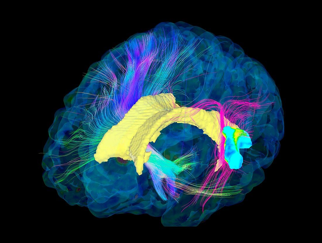
| Brain fibres. 3D diffusion tensor imaging (DTI) magnetic resonance imaging (MRI) scan of a selection of nerve pathways (blue,pink,green) in the brain. The front of the brain is at left. The ventricles are yellow and the brain's cortical surface is also shown. The fibres in the right hemisphere (upper left) are part of the motor system. In the left hemisphere (lower right) are functionally active areas and connections (pink) that go though them. Diffusion tensor imaging measures the direction of water diffusion,which in the brain reveals the orientation of nerve fibres. The technique is also known as tractography,with the resulting image known as a tractogram | |
| Lizenzart: | Lizenzpflichtig |
| Credit: | Science Photo Library / Sherbrooke Connectivity Imaging Lab |
| Bildgröße: | 3425 px × 2587 px |
| Modell-Rechte: | nicht erforderlich |
| Eigentums-Rechte: | nicht erforderlich |
| Restrictions: | - |
Preise für dieses Bild ab 15 €
Universitäten & Organisationen
(Informationsmaterial Digital, Informationsmaterial Print, Lehrmaterial Digital etc.)
ab 15 €
Redaktionell
(Bücher, Bücher: Sach- und Fachliteratur, Digitale Medien (redaktionell) etc.)
ab 30 €
Werbung
(Anzeigen, Aussenwerbung, Digitale Medien, Fernsehwerbung, Karten, Werbemittel, Zeitschriften etc.)
ab 55 €
Handelsprodukte
(bedruckte Textilie, Kalender, Postkarte, Grußkarte, Verpackung etc.)
ab 75 €
Pauschalpreise
Rechtepakete für die unbeschränkte Bildnutzung in Print oder Online
ab 495 €
Keywords
- 3D,
- Anatomie,
- anatomisch,
- Ballaststoff,
- Biologie,
- biologisch,
- cerebral,
- Dreidimensional,
- dti,
- dti Scan,
- farbig,
- Fasern,
- Gehirn,
- gesund,
- Großhirn,
- Hirnscan,
- Magnetresonanztomografie,
- Medizin,
- medizinisch,
- menschlicher Körper,
- MRI,
- MRT-Untersuchung,
- Nerv,
- Nerven,
- Nerventrakt,
- Neuroimaging,
- Neurologie,
- neurologisch,
- normal,
- schwarzer Hintergrund,
- Struktur,
- strukturell,
- Ventrikel,
- Weg,
- Wege,
- weiße Substanz,
- zentrales Nervensystem