Structure of the cochlea,artwork
Bildnummer 11644581
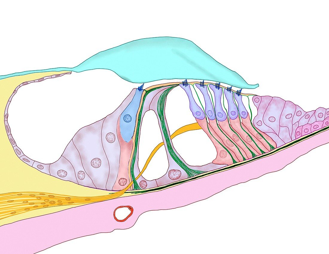
| Structure of the cochlea. Computer artwork of a section through the organ of Corti,the auditory sense organ that lines the spiral of the cochlea in the inner ear. This structure contains rows of hair cells (blue): outer hair cells (right) and inner hair cells (left). On top of these cells are stereocilia,which touch the tectorial membrane (green,top) and detect tiny movements in the membrane. These movements are as a result of sound-induced pressures in the inner ear fluids. The hair cells translate these movements into electrical impulses,which travel down the nerve fibres (orange) to the brain,where they are deciphered as sound | |
| Lizenzart: | Lizenzpflichtig |
| Credit: | Science Photo Library / Veisland, Bo |
| Bildgröße: | 3377 px × 2606 px |
| Modell-Rechte: | nicht erforderlich |
| Restrictions: | - |
Preise für dieses Bild ab 15 €
Universitäten & Organisationen
(Informationsmaterial Digital, Informationsmaterial Print, Lehrmaterial Digital etc.)
ab 15 €
Redaktionell
(Bücher, Bücher: Sach- und Fachliteratur, Digitale Medien (redaktionell) etc.)
ab 30 €
Werbung
(Anzeigen, Aussenwerbung, Digitale Medien, Fernsehwerbung, Karten, Werbemittel, Zeitschriften etc.)
ab 55 €
Handelsprodukte
(bedruckte Textilie, Kalender, Postkarte, Grußkarte, Verpackung etc.)
ab 75 €
Pauschalpreise
Rechtepakete für die unbeschränkte Bildnutzung in Print oder Online
ab 495 €
Keywords
- Anatomie,
- anatomisch,
- aural,
- Biologie,
- biologisch,
- Corti-Organ,
- Fasern,
- Feststellung,
- gesund,
- Hören,
- Illustration,
- Innenohr,
- Klang,
- Kunstwerk,
- menschlicher Körper,
- Nerven,
- Nervensystem,
- Neurologie,
- neurologisch,
- normal,
- Sektion,
- sektioniert,
- Sinn,
- Sinne,
- Struktur,
- Strukturen,
- System,
- Tektorialmembran,
- Tunnel,
- Übermittlung,
- Übertragung,
- weißer Hintergrund,
- Zelle,
- Zellen