Hip fracture reduction,3D CT scan
Bildnummer 11643225
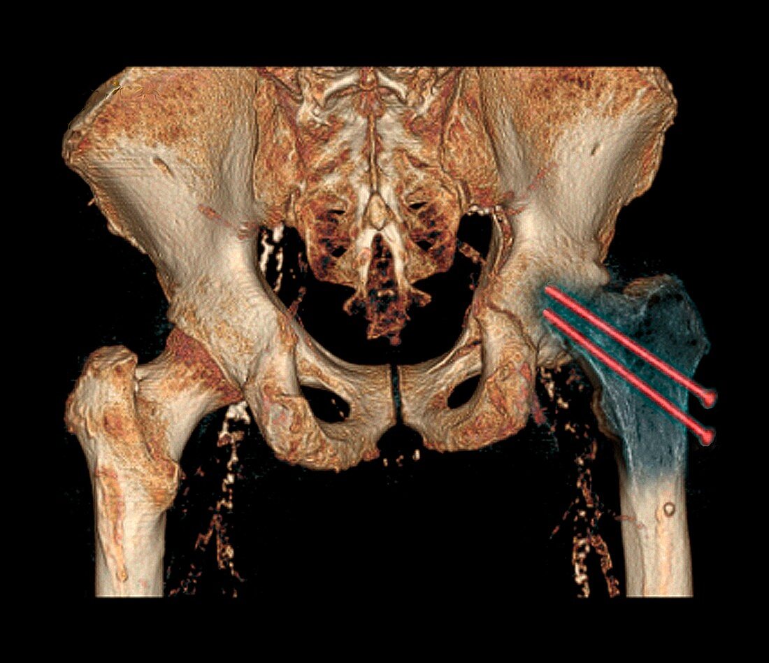
| Hip fracture reduction. Coloured 3D computed tomography (CT) scan of the pelvis of an 81-year-old patient with a fracture to the neck of the right femur (thigh bone). Here,the fracture has been reduced (realigned and fixed) using two metal rods (centre right) | |
| Lizenzart: | Lizenzpflichtig |
| Credit: | Science Photo Library / Zephyr |
| Bildgröße: | 3791 px × 3266 px |
| Modell-Rechte: | nicht erforderlich |
| Restrictions: | - |
Preise für dieses Bild ab 15 €
Universitäten & Organisationen
(Informationsmaterial Digital, Informationsmaterial Print, Lehrmaterial Digital etc.)
ab 15 €
Redaktionell
(Bücher, Bücher: Sach- und Fachliteratur, Digitale Medien (redaktionell) etc.)
ab 30 €
Werbung
(Anzeigen, Aussenwerbung, Digitale Medien, Fernsehwerbung, Karten, Werbemittel, Zeitschriften etc.)
ab 55 €
Handelsprodukte
(bedruckte Textilie, Kalender, Postkarte, Grußkarte, Verpackung etc.)
ab 75 €
Pauschalpreise
Rechtepakete für die unbeschränkte Bildnutzung in Print oder Online
ab 495 €
Keywords
- 3-dimensional,
- 3D,
- abnormal,
- achtziger Jahre,
- Anatomie,
- anatomisch,
- ausgerichtet,
- ausgeschnitten,
- Ausschnitte,
- Becken,
- Behandlung,
- Biologie,
- biologisch,
- Computertomographie,
- ct,
- diagnostische Bildgebung,
- Dreidimensional,
- farbig,
- Femur,
- festgesteckt,
- Fraktur,
- frakturiert,
- gefärbt,
- Gelenk,
- Gesundheitswesen,
- Hüfte,
- Joint,
- kaputt,
- Knochen,
- Kondition,
- Medizin,
- medizinisch,
- menschlicher Körper,
- Nadel,
- Orthopädie,
- orthopädisch,
- pelvin,
- Radiographie,
- Radiologie,
- radiologisch,
- Reduziert,
- Reduzierung,
- Rheumatologie,
- Scan,
- schwarzer Hintergrund,
- Stange,
- Stangen,
- Störung,
- ungesund,
- Unterbrechung