Seminiferous tubule,TEM
Bildnummer 11642153
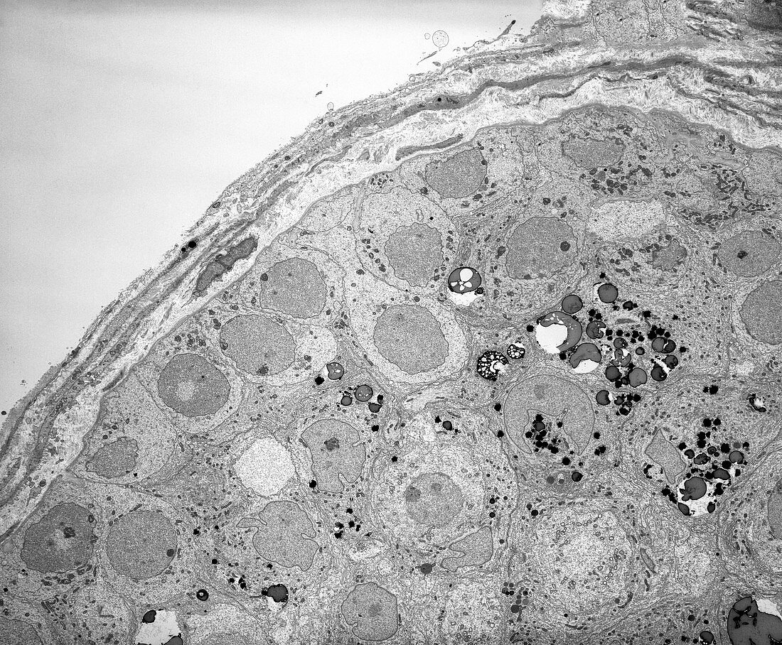
| Seminiferous tubule. Transmission electron micrograph (TEM) of a section through the epithelium of a seminiferous tubule in a human testis (testicle). This is the site of spermatogenesis,the production and maturation of spermatozoa (sperm cells). Seen here are spermatogonia germ cells resting on the basement membrane. Several Sertoli cell nuclei of irregular shape are located just above the spermatogonia. The numerous scattered circular inclusions are lipid droplets found in Sertoli cell cytoplasm. The tissue external to the epithelium is the peritubular tissue,which provides structural support for the tubule. Magnification: x1,000 when printed 10 centimetres wide | |
| Lizenzart: | Lizenzpflichtig |
| Credit: | Science Photo Library / Microscape |
| Bildgröße: | 4638 px × 3828 px |
| Modell-Rechte: | nicht erforderlich |
| Restrictions: | - |
Preise für dieses Bild ab 15 €
Universitäten & Organisationen
(Informationsmaterial Digital, Informationsmaterial Print, Lehrmaterial Digital etc.)
ab 15 €
Redaktionell
(Bücher, Bücher: Sach- und Fachliteratur, Digitale Medien (redaktionell) etc.)
ab 30 €
Werbung
(Anzeigen, Aussenwerbung, Digitale Medien, Fernsehwerbung, Karten, Werbemittel, Zeitschriften etc.)
ab 55 €
Handelsprodukte
(bedruckte Textilie, Kalender, Postkarte, Grußkarte, Verpackung etc.)
ab 75 €
Pauschalpreise
Rechtepakete für die unbeschränkte Bildnutzung in Print oder Online
ab 495 €
Keywords
- Biologie,
- biologisch,
- Bühne,
- Einfarbig,
- Epithel,
- Fortpflanzungssystem,
- Gewebe,
- Histologie,
- histologisch,
- Hoden,
- Männlich,
- Organelle,
- Organellen,
- Reproduktion,
- Samenkanälchen,
- schwarz und weiß,
- Sektion,
- sektioniert,
- Sertoli-Zellen,
- Sperma,
- Spermatozoen,
- Struktur,
- tem,
- Transmissionselektronenmikroskop,
- transmissionselektronenmikroskopische Aufnahme,
- Zellbilogie,
- Zelle,
- Zellen,
- Zytologie,
- Zytologisch