Human spermatid,TEM
Bildnummer 11642143
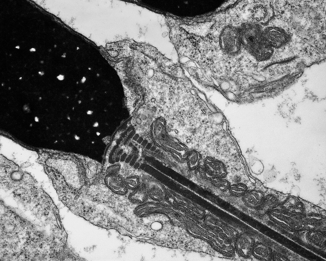
| Human spermatid. Transmission electron micrograph (TEM) of a section through an elongating spermatid in the human testis (testicle),showing the densely compacted nucleus (black,upper left) and part of the tail (flagellum,centre to bottom right). Spermatids mature into spermatozoa (sperm cells). The flagellum is anchored to the spermatid nucleus by striated protein segments. Extending from this are two long columns (black lines) of proteins comprising part of the nine outer dense fibres of the flagellum. Mitochondria form coiled helices around the tail extending as far as the middle piece of the flagellum,and supply the energy needed for sperm motility. Magnification: x25,000 when printed 10 centimetres wide | |
| Lizenzart: | Lizenzpflichtig |
| Credit: | Science Photo Library / Microscape |
| Bildgröße: | 3316 px × 2656 px |
| Modell-Rechte: | nicht erforderlich |
| Restrictions: | - |
Preise für dieses Bild ab 15 €
Universitäten & Organisationen
(Informationsmaterial Digital, Informationsmaterial Print, Lehrmaterial Digital etc.)
ab 15 €
Redaktionell
(Bücher, Bücher: Sach- und Fachliteratur, Digitale Medien (redaktionell) etc.)
ab 30 €
Werbung
(Anzeigen, Aussenwerbung, Digitale Medien, Fernsehwerbung, Karten, Werbemittel, Zeitschriften etc.)
ab 55 €
Handelsprodukte
(bedruckte Textilie, Kalender, Postkarte, Grußkarte, Verpackung etc.)
ab 75 €
Pauschalpreise
Rechtepakete für die unbeschränkte Bildnutzung in Print oder Online
ab 495 €
Keywords
- Atomkern,
- Axonem,
- Ballaststoff,
- Biologie,
- biologisch,
- Bühne,
- Close-up,
- Detail,
- Einfarbig,
- Eiweiß,
- Fasern,
- Filament,
- Filamente,
- Fortpflanzungssystem,
- Geißel,
- Gewebe,
- Histologie,
- histologisch,
- Hoden,
- Männlich,
- Mitochondrien,
- Mitochondrion,
- Organelle,
- Organellen,
- Proteine,
- Reproduktion,
- Schwanz,
- schwarz und weiß,
- Sektion,
- sektioniert,
- Sperma,
- Spermatozoen,
- Struktur,
- tem,
- Transmissionselektronenmikroskop,
- transmissionselektronenmikroskopische Aufnahme,
- Zellbilogie,
- Zelle,
- Zellen,
- Zytologie,
- Zytologisch