Testicular cancer,TEM
Bildnummer 11642142
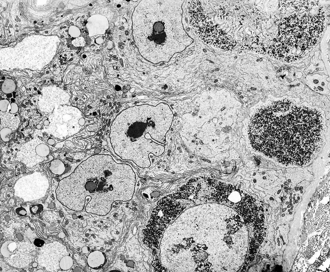
| Testicular cancer. Transmission electron micrograph (TEM) of a section through a testis (testicle),showing neoplastic germ cells (dark,circular),the precursors of testicular cancer. The cells are characteristically round and commonly found next to the basal lamina of the seminiferous tubule's epithelium. Above these premalignant cells are three Sertoli cell nuclei but other germ cells are absent. Also termed carcinoma-in-situ (CIS),the abnormal germ cells often have large amounts of glycogen (black dots) in the cytoplasm,as seen here. Magnification x2,000 when printed 10 centimetres wide | |
| Lizenzart: | Lizenzpflichtig |
| Credit: | Science Photo Library / Microscape |
| Bildgröße: | 4608 px × 3793 px |
| Modell-Rechte: | nicht erforderlich |
| Restrictions: | - |
Preise für dieses Bild ab 15 €
Universitäten & Organisationen
(Informationsmaterial Digital, Informationsmaterial Print, Lehrmaterial Digital etc.)
ab 15 €
Redaktionell
(Bücher, Bücher: Sach- und Fachliteratur, Digitale Medien (redaktionell) etc.)
ab 30 €
Werbung
(Anzeigen, Aussenwerbung, Digitale Medien, Fernsehwerbung, Karten, Werbemittel, Zeitschriften etc.)
ab 55 €
Handelsprodukte
(bedruckte Textilie, Kalender, Postkarte, Grußkarte, Verpackung etc.)
ab 75 €
Pauschalpreise
Rechtepakete für die unbeschränkte Bildnutzung in Print oder Online
ab 495 €
Keywords
- abnormal,
- Biologie,
- biologisch,
- Cis,
- Einfarbig,
- Epithel,
- epithelial,
- Fortpflanzungssystem,
- Gewebe,
- Histologie,
- histologisch,
- Hoden,
- Krebs,
- krebsartig,
- maligne,
- Männlich,
- Medizin,
- medizinisch,
- menschlicher Körper,
- Neoplasma,
- Onkologie,
- onkologisch,
- Organelle,
- Organellen,
- Samenkanälchen,
- schwarz und weiß,
- Sektion,
- sektioniert,
- Struktur,
- tem,
- Transmissionselektronenmikroskop,
- transmissionselektronenmikroskopische Aufnahme,
- Tumor,
- ungesund,
- Zellbilogie,
- Zelle,
- Zellen,
- Zytologie,
- Zytologisch