Kidney anatomy,1825 artwork
Bildnummer 11639263
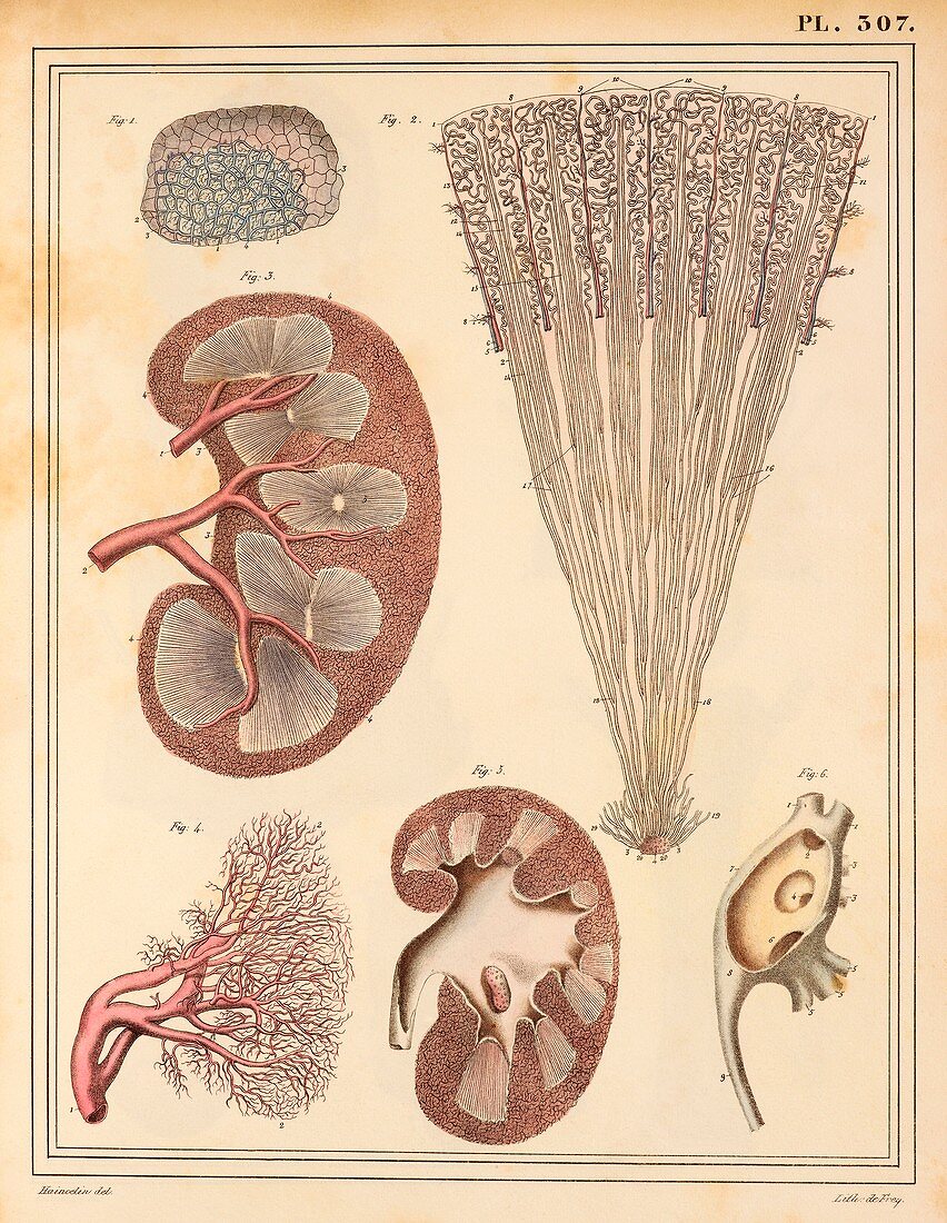
| Kidney anatomy. Artworks of: superficial renal veins (blue,upper left); renal tubules (top right); arteries (red) in sectioned kidney (centre left); renal vessels (wax injection,lower left); ureter (white) in sectioned kidney (bottom centre); kidney stones in ureter (lower right). This anatomical artwork is plate 307 from volume 5 of 'Manuel d'anatomie descriptive du corps humain' (1825). This 5-volume anatomy atlas was produced by French physician and surgeon Jules Germain Cloquet (1790-1883). The illustrations were by Haincelin. Volume 5 illustrated the anatomy of the digestive,secretory and reproductive organs | |
| Lizenzart: | Lizenzpflichtig |
| Credit: | Science Photo Library |
| Bildgröße: | 3860 px × 4984 px |
| Modell-Rechte: | nicht erforderlich |
| Eigentums-Rechte: | nicht erforderlich |
| Restrictions: | - |
Preise für dieses Bild ab 15 €
Universitäten & Organisationen
(Informationsmaterial Digital, Informationsmaterial Print, Lehrmaterial Digital etc.)
ab 15 €
Redaktionell
(Bücher, Bücher: Sach- und Fachliteratur, Digitale Medien (redaktionell) etc.)
ab 30 €
Werbung
(Anzeigen, Aussenwerbung, Digitale Medien, Fernsehwerbung, Karten, Werbemittel, Zeitschriften etc.)
ab 55 €
Handelsprodukte
(bedruckte Textilie, Kalender, Postkarte, Grußkarte, Verpackung etc.)
ab 75 €
Pauschalpreise
Rechtepakete für die unbeschränkte Bildnutzung in Print oder Online
ab 495 €
Keywords
- 1800er Jahre,
- 19. Jahrhundert,
- Abdomen,
- Anatomie,
- anatomische Darstellung,
- Arterien,
- aufgegliedert,
- Cortex,
- europäisch,
- Französisch,
- Geschichte,
- gesund,
- Harnsystem,
- Histologie,
- histologisch,
- historisch,
- Illustration,
- Jules Germain Cloquet,
- Kunstwerk,
- Mark,
- Medizin,
- medizinisch,
- menschlicher Körper,
- Niere,
- Nieren-,
- Nierenstein,
- normal,
- Organ,
- Organe,
- Sektion,
- sektioniert,
- Steine,
- trivial,
- Venen