Heart chamber wall defect,artwork
Bildnummer 11637289
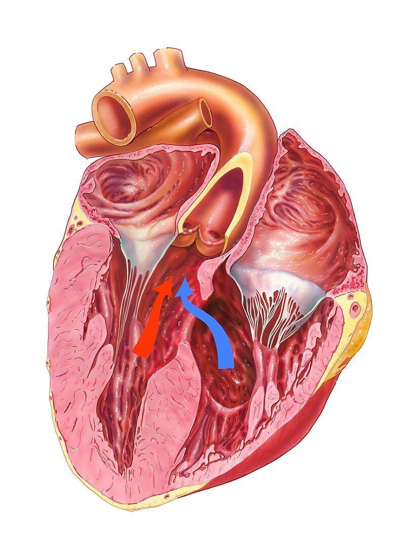
| Heart chamber wall defect. Artwork of a posterior (from behind) view of a defective heart,sectioned to show the internal anatomy. The defect is a hole in the wall (septum) between the heart's two ventricles,known as an ventricular septal defect (VSD). This results in deoxygenated blood (blue arrow) from the right side of the heart mixing with oxygenated blood (red arrow) from the left side of the heart. This can lead to pulmonary hypertension and breathlessness. VSD is usually a congenital defect,present at birth. It can close and heal without intervention,though in severe cases surgery may be needed to repair the defect | |
| Lizenzart: | Lizenzpflichtig |
| Credit: | Science Photo Library / Veisland, Bo |
| Bildgröße: | 3623 px × 4831 px |
| Modell-Rechte: | nicht erforderlich |
| Eigentums-Rechte: | nicht erforderlich |
| Restrictions: | - |
Preise für dieses Bild ab 15 €
Universitäten & Organisationen
(Informationsmaterial Digital, Informationsmaterial Print, Lehrmaterial Digital etc.)
ab 15 €
Redaktionell
(Bücher, Bücher: Sach- und Fachliteratur, Digitale Medien (redaktionell) etc.)
ab 30 €
Werbung
(Anzeigen, Aussenwerbung, Digitale Medien, Fernsehwerbung, Karten, Werbemittel, Zeitschriften etc.)
ab 55 €
Handelsprodukte
(bedruckte Textilie, Kalender, Postkarte, Grußkarte, Verpackung etc.)
ab 75 €
Pauschalpreise
Rechtepakete für die unbeschränkte Bildnutzung in Print oder Online
ab 495 €