Hip anatomy,artwork
Bildnummer 11629737
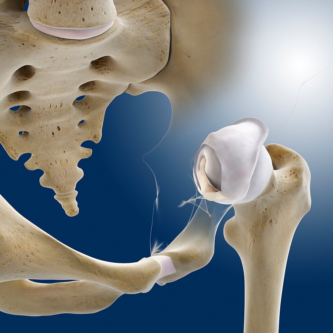
| Hip anatomy. Computer artwork of the head of the left femur (centre right) articulating with the left-hand side of the pelvis to form the hip joint. The bones of the left side of the pelvis are semi-transparent here to show details of the head of the femur. The femoral head and pelvic socket include hyaline cartilage (white) and synovial membrane (white) and fluid. Both cartilage and fluid help to lubricate the joint. At the centre of femoral head is its ligament (ligamentum capitis femoris,fibrous white). The pelvic girdle is at lower centre and the coccyx (tailbone of the spine) is at upper left | |
| Lizenzart: | Lizenzpflichtig |
| Credit: | Science Photo Library / Springer Medizin |
| Bildgröße: | 4180 px × 4180 px |
| Modell-Rechte: | nicht erforderlich |
| Eigentums-Rechte: | nicht erforderlich |
| Restrictions: | - |
Preise für dieses Bild ab 15 €
Universitäten & Organisationen
(Informationsmaterial Digital, Informationsmaterial Print, Lehrmaterial Digital etc.)
ab 15 €
Redaktionell
(Bücher, Bücher: Sach- und Fachliteratur, Digitale Medien (redaktionell) etc.)
ab 30 €
Werbung
(Anzeigen, Aussenwerbung, Digitale Medien, Fernsehwerbung, Karten, Werbemittel, Zeitschriften etc.)
ab 55 €
Handelsprodukte
(bedruckte Textilie, Kalender, Postkarte, Grußkarte, Verpackung etc.)
ab 75 €
Pauschalpreise
Rechtepakete für die unbeschränkte Bildnutzung in Print oder Online
ab 495 €
Keywords
- Anatomie,
- anatomisch,
- Arthrologie,
- Band,
- Bänder,
- Becken,
- Bein,
- Beine,
- Bewegungsapparat,
- Bindegewebe,
- Biologie,
- biologisch,
- Femur,
- Fluid,
- Gelenk,
- Gelenke,
- gesund,
- Hüfte,
- Hüften,
- Hüftknochen,
- Illustration,
- Joint,
- Kapsel,
- Knochen,
- Knorpel,
- Kreuzbein,
- Kunstwerk,
- menschlicher Körper,
- normal,
- Oberschenkelknochen,
- Osteologie,
- pelvin,
- Schenkel,
- Skelett,
- Skelett-,
- Steißbein,
- Synovialmembran,
- untere Gliedmaßen,
- unterer Rücken,
- Wirbelsäule