Internal heart anatomy,3D CT scan
Bildnummer 11628719
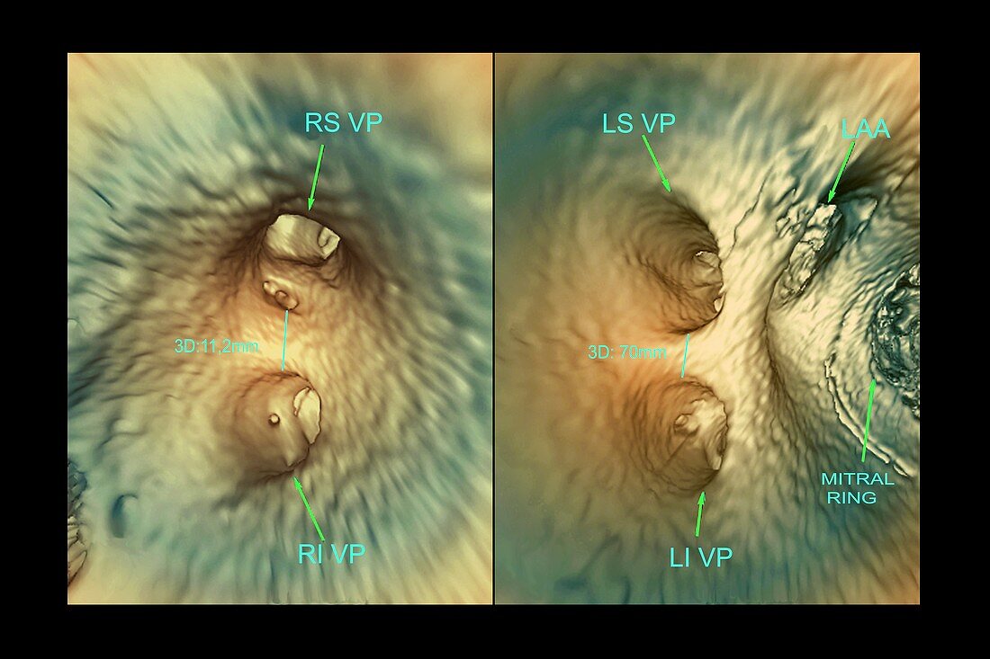
| Internal heart anatomy,coloured 3D computed tomography (CT) scans. Interior of the left atrium of the heart of a 57-year-old patient with atrial fibrillation (irregular heartbeat),showing the openings (ostia) of the right (R) and left (L) inferior (I) and superior (S) pulmonary veins (VP). This image was taken to assess the existence or absence of blood clots (thrombi) in the heart chambers | |
| Lizenzart: | Lizenzpflichtig |
| Credit: | Science Photo Library / Zephyr |
| Bildgröße: | 4589 px × 3058 px |
| Modell-Rechte: | nicht erforderlich |
| Restrictions: | - |
Preise für dieses Bild ab 15 €
Universitäten & Organisationen
(Informationsmaterial Digital, Informationsmaterial Print, Lehrmaterial Digital etc.)
ab 15 €
Redaktionell
(Bücher, Bücher: Sach- und Fachliteratur, Digitale Medien (redaktionell) etc.)
ab 30 €
Werbung
(Anzeigen, Aussenwerbung, Digitale Medien, Fernsehwerbung, Karten, Werbemittel, Zeitschriften etc.)
ab 55 €
Handelsprodukte
(bedruckte Textilie, Kalender, Postkarte, Grußkarte, Verpackung etc.)
ab 75 €
Pauschalpreise
Rechtepakete für die unbeschränkte Bildnutzung in Print oder Online
ab 495 €
Keywords
- 3-dimensional,
- 3D,
- 50er Jahre,
- Anatomie,
- anatomisch,
- Atrial,
- Atrium,
- beschriftet,
- Computertomographie,
- ct,
- Diagnose,
- diagnostische Bildgebung,
- Dreidimensional,
- Etikette,
- Etiketten,
- Farbig,
- Fünfziger Jahre,
- gefärbt,
- Herz,
- Herzkreislaufsystem,
- intern,
- Kammer,
- Kreislauf,
- linkes Atrium,
- links,
- Lungenvene,
- Organ,
- Ostia,
- Radiographie,
- Radiologie,
- radiologisch,
- Recht,
- Röntgen,
- Scan,
- Struktur,
- überlegen,
- vaskulär,
- Venen