Neck blood vessels in stroke patient
Bildnummer 11628702
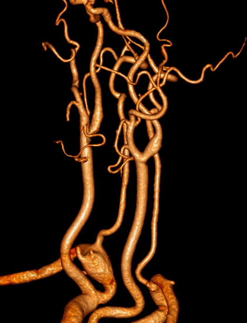
| Neck blood vessels in stroke patient. Coloured 3D computed tomography (CT) angiogram of the blood vessels in the neck of a 60-year-old patient with a history of transient ischaemic attacks (mini strokes). These are the blood vessels that supply the brain. The vertebral arteries (thin,lower centre left and right) are healthy. However,the carotid arteries (thick,lower centre left and right) show abnormal thickness,with probable presence of atheromatous plaques (lumpy areas) in the area of the carotid bifurcations (forked areas,centre) - where the common carotid arteries divide into the internal and external carotid arteries | |
| Lizenzart: | Lizenzpflichtig |
| Credit: | Science Photo Library / Zephyr |
| Bildgröße: | 3655 px × 4779 px |
| Modell-Rechte: | nicht erforderlich |
| Restrictions: | - |
Preise für dieses Bild ab 15 €
Universitäten & Organisationen
(Informationsmaterial Digital, Informationsmaterial Print, Lehrmaterial Digital etc.)
ab 15 €
Redaktionell
(Bücher, Bücher: Sach- und Fachliteratur, Digitale Medien (redaktionell) etc.)
ab 30 €
Werbung
(Anzeigen, Aussenwerbung, Digitale Medien, Fernsehwerbung, Karten, Werbemittel, Zeitschriften etc.)
ab 55 €
Handelsprodukte
(bedruckte Textilie, Kalender, Postkarte, Grußkarte, Verpackung etc.)
ab 75 €
Pauschalpreise
Rechtepakete für die unbeschränkte Bildnutzung in Print oder Online
ab 495 €
Keywords
- 3-dimensional,
- 3D,
- 60er Jahre,
- abnormal,
- Alt,
- älter,
- Angiografie,
- Angiogramm,
- Arterie,
- arteriell,
- Arterien,
- Atherom,
- Blutgefäß,
- Blutversorgung,
- Computertomographie,
- ct,
- Dreidimensional,
- extern,
- Gefäße,
- Gehirn,
- Hals,
- Herzkreislaufsystem,
- intern,
- Kofferraum,
- Kopf,
- Medizin,
- medizinisch,
- menschlicher Körper,
- Mini-Schlaganfall,
- Röntgen,
- Scan,
- Scanner,
- schwarzer Hintergrund,
- sechziger Jahre,
- ungesund,
- vertebral