MEG and fMRI Brain Scans
Bildnummer 11627765
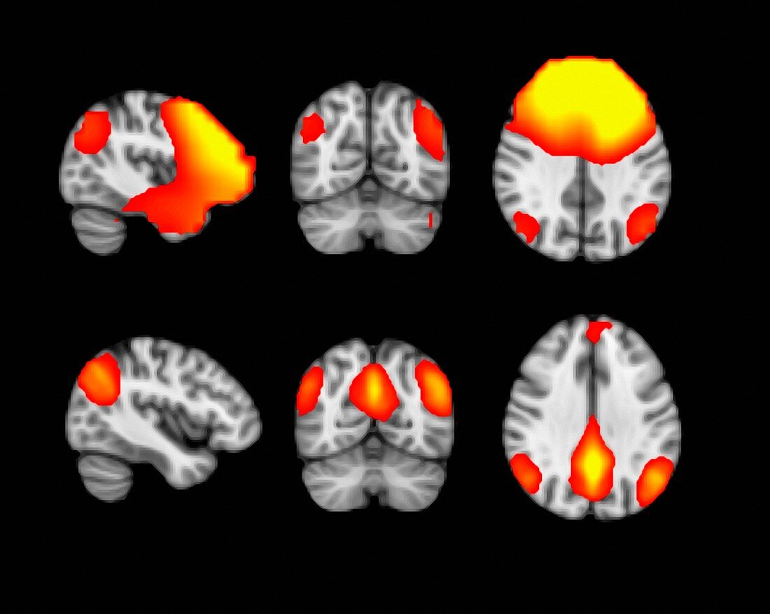
| Magnetoencephalography (MEG) scans (top row) of slices of a human brain,and comparison scans using Functional Magnetic Resonance Imaging (fMRI). Both sets of scans show network activity in the resting or 'default-mode' state. This network is only found at rest and disappears when the subject is given a task. The 'default-mode' network and its connection to various neurological diseases and disorders is currently being intensively researched.The MEG scanner images the magnetic fields produced by the electrical currents resulting from neural activity within the brain. fMRI scanning measures the changes in blood flow resulting from the neural activity caused by brain stimulation or tasking. On both types of image the gradient of activity is from areas of low (red) to high (yellow) | |
| Lizenzart: | Lizenzpflichtig |
| Credit: | Science Photo Library / HENRY LUCKHOO / JAMES KING-HOLMES |
| Bildgröße: | 3320 px × 2646 px |
| Modell-Rechte: | nicht erforderlich |
| Eigentums-Rechte: | nicht erforderlich |
| Restrictions: | - |
Preise für dieses Bild ab 15 €
Universitäten & Organisationen
(Informationsmaterial Digital, Informationsmaterial Print, Lehrmaterial Digital etc.)
ab 15 €
Redaktionell
(Bücher, Bücher: Sach- und Fachliteratur, Digitale Medien (redaktionell) etc.)
ab 30 €
Werbung
(Anzeigen, Aussenwerbung, Digitale Medien, Fernsehwerbung, Karten, Werbemittel, Zeitschriften etc.)
ab 55 €
Handelsprodukte
(bedruckte Textilie, Kalender, Postkarte, Grußkarte, Verpackung etc.)
ab 75 €
Pauschalpreise
Rechtepakete für die unbeschränkte Bildnutzung in Print oder Online
ab 495 €