Scanning human fossil thigh bones
Bildnummer 11623636
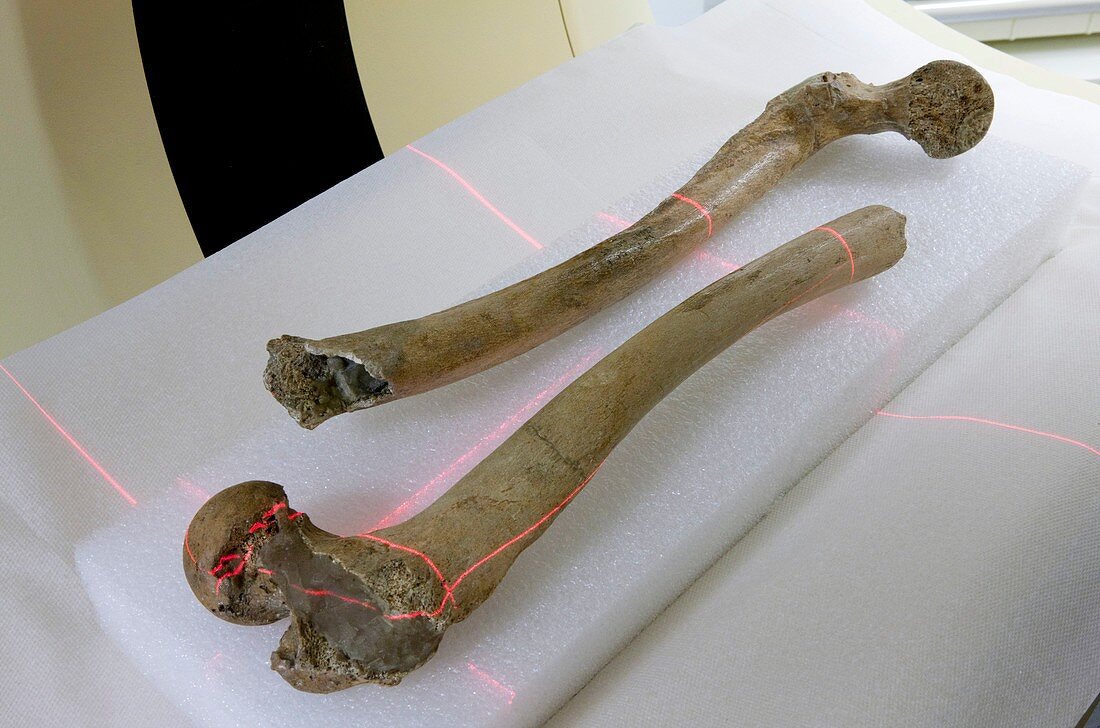
| Scanning human fossil thigh bones. Medical scanner being used to scan specimens of human fossil thigh bones (femurs) that include the femoral head and neck. Scans such as this are carried out to examine the internal and detailed anatomy of the fossils,and to help construct detailed 3D models. The structure and development of this bone,which varies with age,can help determine the age of the individual. The red lights are laser guide lines. These specimens are from the Musee de l'Homme (Museum of Man),Paris,France,one of the departments of the French National Museum of Natural History (MNHN). Photographed in Paris,in 2010 | |
| Lizenzart: | Lizenzpflichtig |
| Credit: | Science Photo Library / Goetgheluck, Pascal |
| Bildgröße: | 5194 px × 3437 px |
| Modell-Rechte: | nicht erforderlich |
| Eigentums-Rechte: | nicht erforderlich |
| Restrictions: | - |
Preise für dieses Bild ab 15 €
Universitäten & Organisationen
(Informationsmaterial Digital, Informationsmaterial Print, Lehrmaterial Digital etc.)
ab 15 €
Redaktionell
(Bücher, Bücher: Sach- und Fachliteratur, Digitale Medien (redaktionell) etc.)
ab 30 €
Werbung
(Anzeigen, Aussenwerbung, Digitale Medien, Fernsehwerbung, Karten, Werbemittel, Zeitschriften etc.)
ab 55 €
Handelsprodukte
(bedruckte Textilie, Kalender, Postkarte, Grußkarte, Verpackung etc.)
ab 75 €
Pauschalpreise
Rechtepakete für die unbeschränkte Bildnutzung in Print oder Online
ab 495 €
Keywords
- 2010,
- 21. Jahrhundert,
- Anthropologie,
- anthropologisch,
- Ausrüstung,
- Duo,
- Europa,
- europäisch,
- evolutionär,
- Femur,
- Forschung,
- Fossil,
- fossiler Mann,
- Fossilien,
- fossilisiert,
- Frankreich,
- Französisch,
- Geschichte,
- historisch,
- Knochen,
- Krankenhaus,
- Licht,
- Linien,
- menschliche Evolution,
- menschliche Fossilien,
- MNHN,
- Museum,
- Museum des Menschen,
- Oberschenkelhals,
- Oberschenkelknochen,
- Paar,
- Paläoanthropologie,
- Paläontologie,
- paläontologisch,
- Paris,
- prähistorisch,
- Probe,
- Scanner,
- Technologie,
- technologisch,
- versteinert,
- Vorgeschichte,
- Zwei