Pad from a dog's foot,light micrograph
Bildnummer 11623305
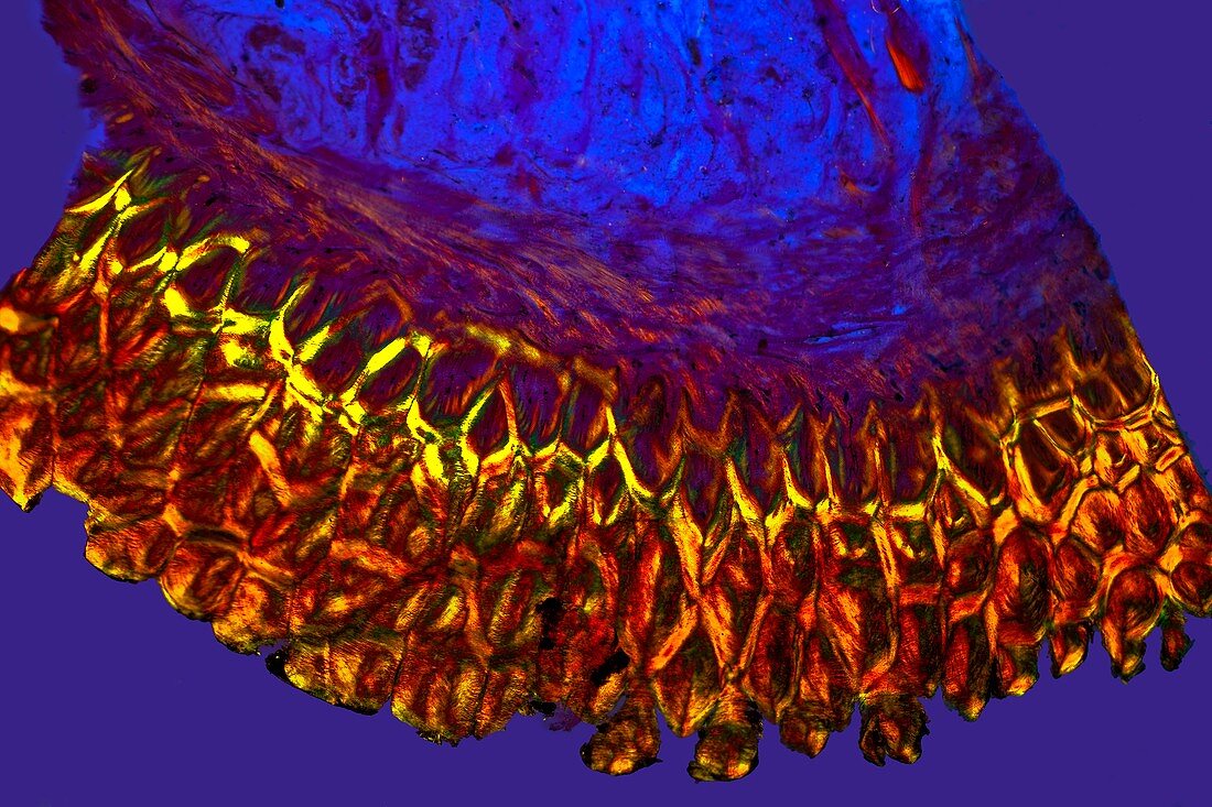
| Pad from a dog's foot. Polarised light micrograph of a section through a pad from a dog's foot. The hard pad originates from the epidermis (surface layer) of the skin. It consists of a thick keratinised layer of stratified squamous epithelium (brown and orange). Beneath this is the dermis (red),which contains fibrous and elastic connective tissue,and the hypodermis (blue),which contains adipose (fatty) tissue | |
| Lizenzart: | Lizenzpflichtig |
| Credit: | Science Photo Library / Wheeler, Dr. Keith |
| Bildgröße: | 5138 px × 3425 px |
| Modell-Rechte: | nicht erforderlich |
| Eigentums-Rechte: | nicht erforderlich |
| Restrictions: | - |
Preise für dieses Bild ab 15 €
Universitäten & Organisationen
(Informationsmaterial Digital, Informationsmaterial Print, Lehrmaterial Digital etc.)
ab 15 €
Redaktionell
(Bücher, Bücher: Sach- und Fachliteratur, Digitale Medien (redaktionell) etc.)
ab 30 €
Werbung
(Anzeigen, Aussenwerbung, Digitale Medien, Fernsehwerbung, Karten, Werbemittel, Zeitschriften etc.)
ab 55 €
Handelsprodukte
(bedruckte Textilie, Kalender, Postkarte, Grußkarte, Verpackung etc.)
ab 75 €
Pauschalpreise
Rechtepakete für die unbeschränkte Bildnutzung in Print oder Online
ab 495 €