Magnetoencephalograph scanning
Bildnummer 11623043
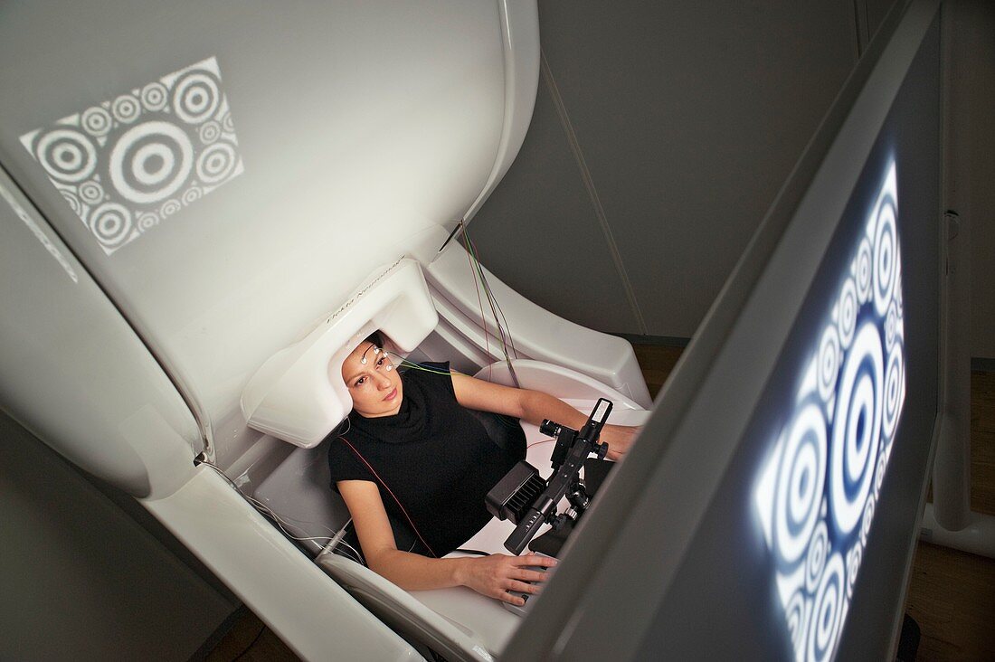
| The brain of a research subject is stimulated by visual images whilst superconducting sensors in a magnetoencephalograph (MEG) records the neural activity. The MEG scanner uses 306 highly sensitive SQUIDs (Superconducting Quantum Interface Devices) which detect the extremely weak magnetic fields and associated gradients generated by neural activity in different parts of the brain. The resulting data is used with other neuro-imaging techniques to investigate psychiatric and neurological disorders such as ADHD,epilepsy and age-related memory and cognitive syndromes such as Alzheimer's and dementia. The eye-tracking device in front of the subject gives data on the gaze related to the brain activity and also relays information on any blinking or head movement which may influence the brain's neural currents. Photographed at the Oxford Centre for Human Brain Activity | |
| Lizenzart: | Lizenzpflichtig |
| Credit: | Science Photo Library / King-Holmes, James |
| Bildgröße: | 5140 px × 3420 px |
| Modell-Rechte: | vorhanden |
| Eigentums-Rechte: | nicht erforderlich |
| Restrictions: | - |
Preise für dieses Bild ab 15 €
Universitäten & Organisationen
(Informationsmaterial Digital, Informationsmaterial Print, Lehrmaterial Digital etc.)
ab 15 €
Redaktionell
(Bücher, Bücher: Sach- und Fachliteratur, Digitale Medien (redaktionell) etc.)
ab 30 €
Werbung
(Anzeigen, Aussenwerbung, Digitale Medien, Fernsehwerbung, Karten, Werbemittel, Zeitschriften etc.)
ab 55 €
Handelsprodukte
(bedruckte Textilie, Kalender, Postkarte, Grußkarte, Verpackung etc.)
ab 75 €
Pauschalpreise
Rechtepakete für die unbeschränkte Bildnutzung in Print oder Online
ab 495 €