Chicken embryo,light micrograph
Bildnummer 11621189
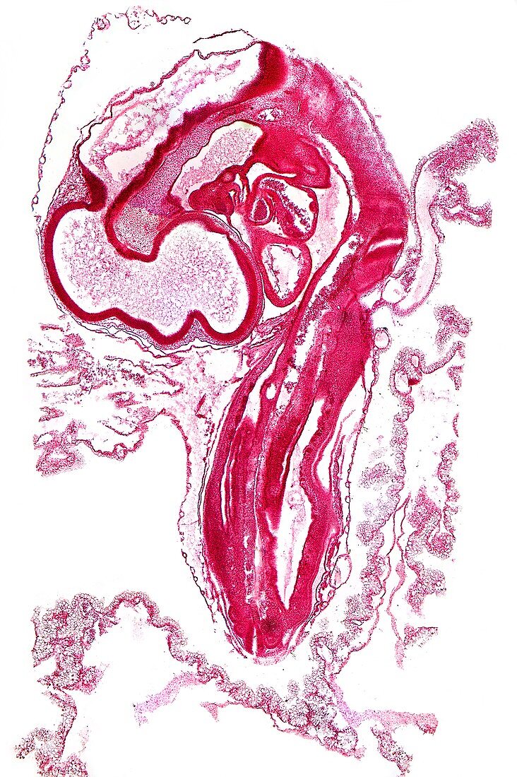
| Chicken embryo. Light micrograph of a longitudinal section through a 3-day-old chicken embryo. The head is at upper left,and includes the brain,fore-brain,mid-brain,and hind-brain. The body's somites (solid red,vertebral segments) go from upper centre to lower centre,with the spinal cord (white tubes) between them. The heart (upper centre) is between the brain and the body. Parts of the yolk arteries and veins are seen surrounding the embryo. Magnification: x13 when printed 10 centimetres tall | |
| Lizenzart: | Lizenzpflichtig |
| Credit: | Science Photo Library / Wheeler, Dr. Keith |
| Bildgröße: | 3411 px × 5125 px |
| Modell-Rechte: | nicht erforderlich |
| Eigentums-Rechte: | nicht erforderlich |
| Restrictions: | - |
Preise für dieses Bild ab 15 €
Universitäten & Organisationen
(Informationsmaterial Digital, Informationsmaterial Print, Lehrmaterial Digital etc.)
ab 15 €
Redaktionell
(Bücher, Bücher: Sach- und Fachliteratur, Digitale Medien (redaktionell) etc.)
ab 30 €
Werbung
(Anzeigen, Aussenwerbung, Digitale Medien, Fernsehwerbung, Karten, Werbemittel, Zeitschriften etc.)
ab 55 €
Handelsprodukte
(bedruckte Textilie, Kalender, Postkarte, Grußkarte, Verpackung etc.)
ab 75 €
Pauschalpreise
Rechtepakete für die unbeschränkte Bildnutzung in Print oder Online
ab 495 €
Keywords
- Anatomie,
- anatomisch,
- Biologie,
- biologisch,
- Ei,
- Eigelb,
- einer,
- Embryo,
- Embryologie,
- Entwicklung,
- Entwicklungsbiologie,
- Fauna,
- Gehirn,
- gesund,
- Hähnchen,
- Herz,
- Histologie,
- histologisch,
- Kopf,
- Körper,
- Längsschnitt,
- Lichtmikroskop,
- lichtmikroskopische Aufnahme,
- Muskel,
- Muskeln,
- Natur,
- normal,
- ornithologisch,
- Rückenmark,
- Rückgrat,
- Sektion,
- sektioniert,
- Single,
- Somiten,
- Tier,
- Tierwelt,
- vertebral,
- Vogel,
- Vogelkunde,
- weißer Hintergrund,
- Wirbelsäulen-,
- Zoologie,
- zoologisch