Pine root,light micrograph
Bildnummer 11619932
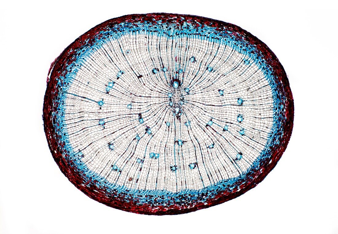
| Pine root. Light micrograph of a section through the root of a pine (Pinus sp.) tree. From outer to inwards: outer layer of peridium (dark red); cortex - made up of parenchyma cells (red); ring of secondary phloem (blue); cambium (blue); secondary xylem - made up of tracheid cells (pink),scattered resin canals (blue,circular),and meullary rays (dark radial lines). The centre of the root is filled with small primary xylem cells (metaxylem,brown). Magnification: x5 when printed 10 centimetres wide | |
| Lizenzart: | Lizenzpflichtig |
| Credit: | Science Photo Library / Wheeler, Dr. Keith |
| Bildgröße: | 5016 px × 3482 px |
| Modell-Rechte: | nicht erforderlich |
| Restrictions: | - |
Preise für dieses Bild ab 15 €
Universitäten & Organisationen
(Informationsmaterial Digital, Informationsmaterial Print, Lehrmaterial Digital etc.)
ab 15 €
Redaktionell
(Bücher, Bücher: Sach- und Fachliteratur, Digitale Medien (redaktionell) etc.)
ab 30 €
Werbung
(Anzeigen, Aussenwerbung, Digitale Medien, Fernsehwerbung, Karten, Werbemittel, Zeitschriften etc.)
ab 55 €
Handelsprodukte
(bedruckte Textilie, Kalender, Postkarte, Grußkarte, Verpackung etc.)
ab 75 €
Pauschalpreise
Rechtepakete für die unbeschränkte Bildnutzung in Print oder Online
ab 495 €
Keywords
- ausgeschnitten,
- Ausschnitte,
- Baum,
- befleckt,
- Biologie,
- biologisch,
- Botanik,
- botanisch,
- Close-up,
- Cortex,
- Detail,
- Gang,
- Gewebe,
- gymnospermen,
- Histologie,
- histologisch,
- Holz,
- Kambium,
- Kiefer,
- Lichtmikroskop,
- lichtmikroskopische Aufnahme,
- Mikroskopie,
- Nacktsamer,
- Parenchym,
- Pflanze,
- Pflanzen,
- primäres Xylem,
- Sektion,
- sektioniert,
- Strahlen,
- Struktur,
- strukturell,
- Strukturen,
- Verfärbung,
- weißer Hintergrund,
- Wurzel,
- Zellbilogie,
- Zelle,
- Zellen,
- Zytologie,
- Zytologisch