Pine stem,light micrograph
Bildnummer 11619922
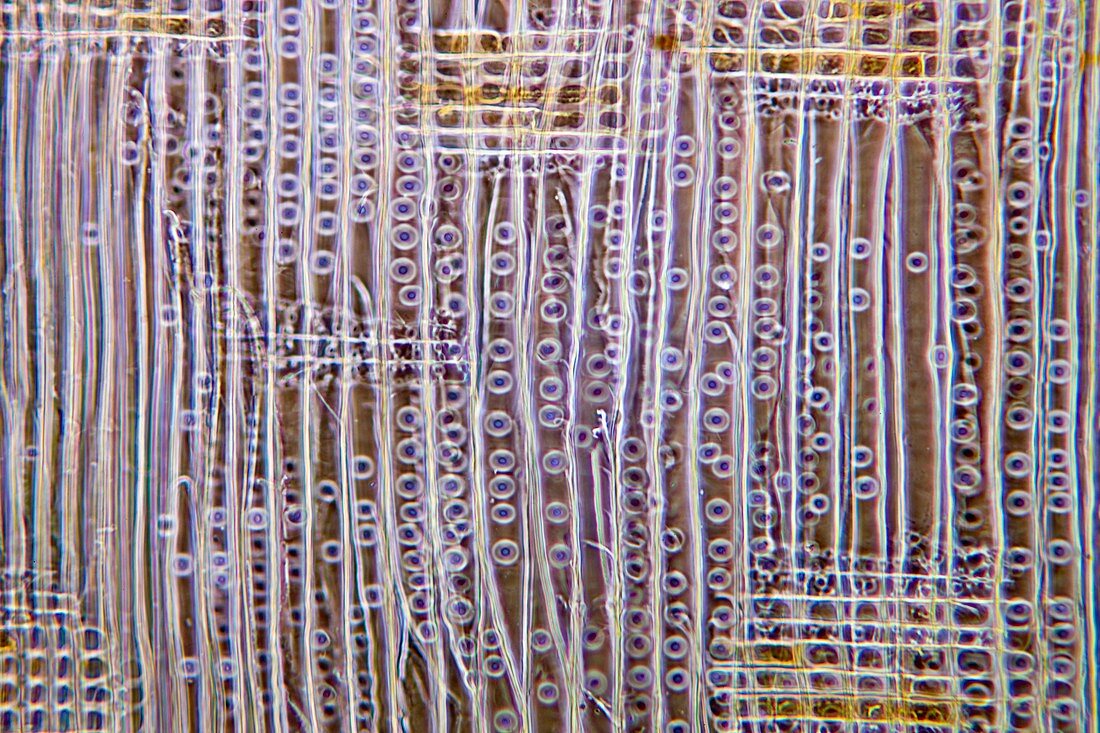
| Pine stem. Light micrograph of a section through a single annual growth ring in the stem of a pine (Pinus sp.) tree,showing xylem tissue. The large thin-walled tracheid cells (white-brown) are of younger spring wood (right of image),whilst smaller thick-walled tracheid cells (also white-brown) are of older autumn wood (left of image). There are thin files of parenchyma and tracheid cells (yellow-white) of the subsidiary medullary rays,which pass food substances radially. Many of the tracheid cell walls have bordered pits (circles) that pass water and minerals side-ways. Magnification: x37 when printed 10 centimetres wide | |
| Lizenzart: | Lizenzpflichtig |
| Credit: | Science Photo Library / Wheeler, Dr. Keith |
| Bildgröße: | 5125 px × 3417 px |
| Modell-Rechte: | nicht erforderlich |
| Restrictions: | - |
Preise für dieses Bild ab 15 €
Universitäten & Organisationen
(Informationsmaterial Digital, Informationsmaterial Print, Lehrmaterial Digital etc.)
ab 15 €
Redaktionell
(Bücher, Bücher: Sach- und Fachliteratur, Digitale Medien (redaktionell) etc.)
ab 30 €
Werbung
(Anzeigen, Aussenwerbung, Digitale Medien, Fernsehwerbung, Karten, Werbemittel, Zeitschriften etc.)
ab 55 €
Handelsprodukte
(bedruckte Textilie, Kalender, Postkarte, Grußkarte, Verpackung etc.)
ab 75 €
Pauschalpreise
Rechtepakete für die unbeschränkte Bildnutzung in Print oder Online
ab 495 €
Keywords
- Alt,
- älter,
- Baum,
- befleckt,
- Biologie,
- biologisch,
- Botanik,
- botanisch,
- Close-up,
- Detail,
- Frühling,
- Gefäß,
- Gefäße,
- Gewebe,
- gymnospermen,
- Herbst,
- Histologie,
- histologisch,
- Holz,
- Jung,
- jünger,
- Kiefer,
- Kofferraum,
- Lichtmikroskop,
- lichtmikroskopische Aufnahme,
- Mikroskopie,
- Nacktsamer,
- Parenchym,
- Pflanze,
- Pflanzen,
- Sektion,
- sektioniert,
- Stengel,
- Strahl,
- Struktur,
- strukturell,
- Strukturen,
- Verfärbung,
- wachsend,
- Zellbilogie,
- Zelle,
- Zellen,
- Zytologie,
- Zytologisch