Lime tree stem,light micrograph
Bildnummer 11619029
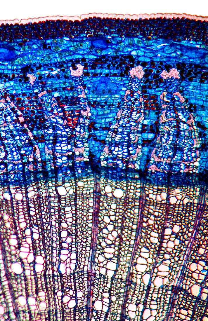
| Lime tree stem. Light micrograph of a section through the stem of a lime tree (Tilia europaea). The outer epidermis has been shed and replaced by a layer of cork (dark red). Under the cork is the outer cortex layer of flexible collenchyma tissue (dark blue and red) and a lower cortex of parenchyma (blue). Next is a ring of phloem (blue) with large sieve tubes,companion cells and fibres (pink). The phloem is broken by primary medullary rays (blue,triangular) and thin subsidiary medullary rays (dark blue). Next is a ring of xylem (light pink) with large files of vessels and small woody parenchyma (dark red-brown). There are two yearly growth rings,which have large vessels laid down in the spring and small vessels in the previous autumn. Magnification: | |
| Lizenzart: | Lizenzpflichtig |
| Credit: | Science Photo Library / Wheeler, Dr. Keith |
| Bildgröße: | 3370 px × 5203 px |
| Modell-Rechte: | nicht erforderlich |
| Restrictions: | - |
Preise für dieses Bild ab 15 €
Universitäten & Organisationen
(Informationsmaterial Digital, Informationsmaterial Print, Lehrmaterial Digital etc.)
ab 15 €
Redaktionell
(Bücher, Bücher: Sach- und Fachliteratur, Digitale Medien (redaktionell) etc.)
ab 30 €
Werbung
(Anzeigen, Aussenwerbung, Digitale Medien, Fernsehwerbung, Karten, Werbemittel, Zeitschriften etc.)
ab 55 €
Handelsprodukte
(bedruckte Textilie, Kalender, Postkarte, Grußkarte, Verpackung etc.)
ab 75 €
Pauschalpreise
Rechtepakete für die unbeschränkte Bildnutzung in Print oder Online
ab 495 €
Keywords
- Angiosperme,
- Angiospermen,
- Ballaststoff,
- befleckt,
- Biologie,
- biologisch,
- Botanik,
- botanisch,
- Close-up,
- Cortex,
- Detail,
- Dikotyle,
- Epidermis,
- Fasern,
- Gefäße,
- Gefäßgewebe,
- Gewebe,
- Histologie,
- histologisch,
- Holz,
- holzig,
- Kork,
- Lichtmikroskop,
- lichtmikroskopische Aufnahme,
- Mikroskopie,
- Parenchym,
- Pflanze,
- Pflanzen,
- Phloem,
- primäres Xylem,
- Ringe,
- Röhren,
- Schicht,
- Schichten,
- Sektion,
- sektioniert,
- Stengel,
- Strahlen,
- Struktur,
- strukturell,
- Strukturen,
- Verfärbung,
- weißer Hintergrund,
- Xylemgefäß,
- Zellbilogie,
- Zelle,
- Zellen,
- Zytologie,
- Zytologisch