Lime tree stem,light micrograph
Bildnummer 11619028
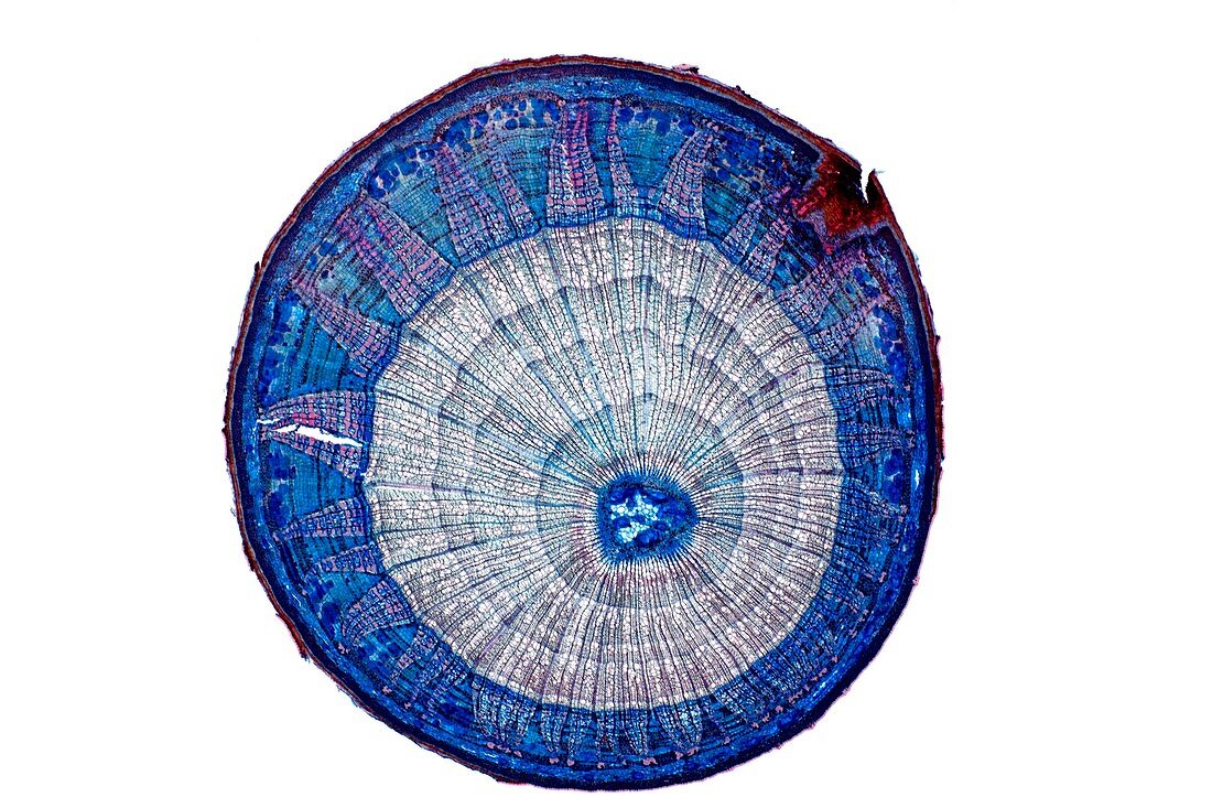
| Lime tree stem. Light micrograph of a section through the stem of a lime tree (Tilia europaea). The outer epidermis has been shed and replaced by a layer of cork (dark red). Under the cork is the outer cortex layer of flexible collenchyma tissue (dark blue and red) and a lower cortex of parenchyma (blue). Next is a ring of phloem (blue) with large sieve tubes,companion cells and fibres (pink). The phloem is broken by primary medullary rays (blue,triangular) and thin subsidiary medullary rays (dark blue). Next is a ring of xylem (pink) with large files of vessels and small woody parenchyma (light blue). The primary xylem (dark blue) and the central pith of parenchyma tissue (light blue) are also shown. Magnification: x5 when printed 10 centimetres | |
| Lizenzart: | Lizenzpflichtig |
| Credit: | Science Photo Library / Wheeler, Dr. Keith |
| Bildgröße: | 5125 px × 3417 px |
| Modell-Rechte: | nicht erforderlich |
| Restrictions: | - |
Preise für dieses Bild ab 15 €
Universitäten & Organisationen
(Informationsmaterial Digital, Informationsmaterial Print, Lehrmaterial Digital etc.)
ab 15 €
Redaktionell
(Bücher, Bücher: Sach- und Fachliteratur, Digitale Medien (redaktionell) etc.)
ab 30 €
Werbung
(Anzeigen, Aussenwerbung, Digitale Medien, Fernsehwerbung, Karten, Werbemittel, Zeitschriften etc.)
ab 55 €
Handelsprodukte
(bedruckte Textilie, Kalender, Postkarte, Grußkarte, Verpackung etc.)
ab 75 €
Pauschalpreise
Rechtepakete für die unbeschränkte Bildnutzung in Print oder Online
ab 495 €
Keywords
- Angiosperme,
- Angiospermen,
- ausgeschnitten,
- Ausschnitte,
- Ballaststoff,
- befleckt,
- Biologie,
- biologisch,
- Botanik,
- botanisch,
- Close-up,
- Cortex,
- Detail,
- Dikotyle,
- Epidermis,
- Fasern,
- Gefäße,
- Gefäßgewebe,
- Gewebe,
- Histologie,
- histologisch,
- Holz,
- holzig,
- Kork,
- Lichtmikroskop,
- lichtmikroskopische Aufnahme,
- Mikroskopie,
- Parenchym,
- Pflanze,
- Pflanzen,
- Phloem,
- primäres Xylem,
- Röhren,
- Schicht,
- Schichten,
- Sektion,
- sektioniert,
- Stengel,
- Strahlen,
- Struktur,
- strukturell,
- Strukturen,
- Verfärbung,
- weißer Hintergrund,
- Xylemgefäß,
- Zellbilogie,
- Zelle,
- Zellen,
- Zytologie,
- Zytologisch