Sage stem,light micrograph
Bildnummer 11619026
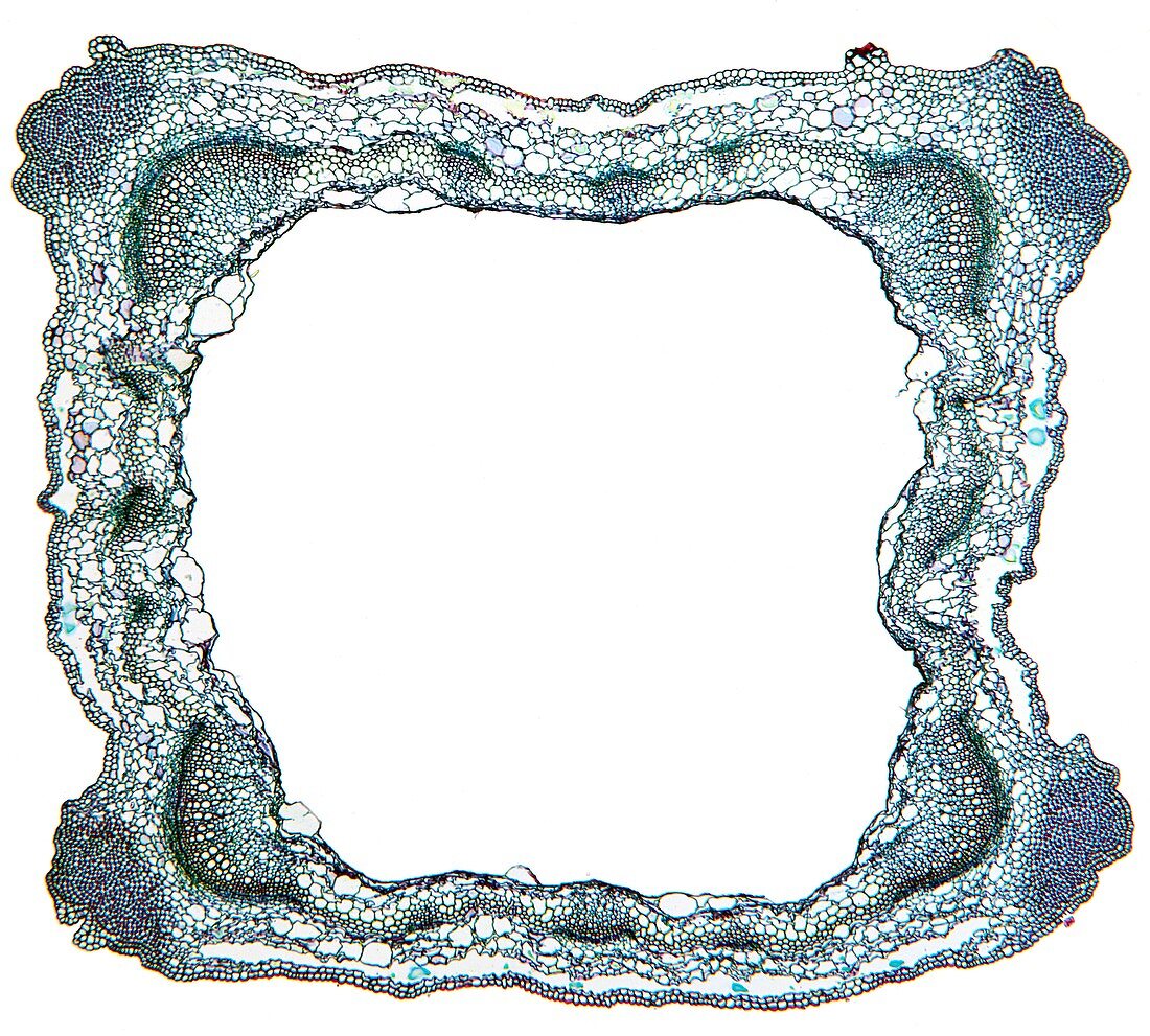
| Sage stem. Light micrograph of a section through a primary stem of a scarlet sage (Salivia splendens) plant. The outer stem is covered with a thin epidermis (green) that contains stomata. Under the epidermis,at the corners,is a layer of flexible collenchyma (blue-red) for support. The cortex is made up of parenchyma cells (round,red). Next are four large and twelve small vascular bundles. Each has an thin outer layer of phloem (green) with sieve plates and companion cells,and an inner xylem (red),with large vessels and small-celled woody parenchyma tissue (green). In-between is a thin layer of cambium. The centre of the stem is a pith of parenchyma (green),most of which has broken,leaving a hollow. Magnification: x5 when printed 10 centimetres | |
| Lizenzart: | Lizenzpflichtig |
| Credit: | Science Photo Library / Wheeler, Dr. Keith |
| Bildgröße: | 4424 px × 3985 px |
| Modell-Rechte: | nicht erforderlich |
| Restrictions: | - |
Preise für dieses Bild ab 15 €
Universitäten & Organisationen
(Informationsmaterial Digital, Informationsmaterial Print, Lehrmaterial Digital etc.)
ab 15 €
Redaktionell
(Bücher, Bücher: Sach- und Fachliteratur, Digitale Medien (redaktionell) etc.)
ab 30 €
Werbung
(Anzeigen, Aussenwerbung, Digitale Medien, Fernsehwerbung, Karten, Werbemittel, Zeitschriften etc.)
ab 55 €
Handelsprodukte
(bedruckte Textilie, Kalender, Postkarte, Grußkarte, Verpackung etc.)
ab 75 €
Pauschalpreise
Rechtepakete für die unbeschränkte Bildnutzung in Print oder Online
ab 495 €
Keywords
- Angiosperme,
- Angiospermen,
- befleckt,
- Biologie,
- biologisch,
- Botanik,
- botanisch,
- Bündel,
- Close-up,
- Cortex,
- Detail,
- Dikotyle,
- Epidermis,
- Gefäß,
- Gefäßband,
- Gefäße,
- Gefäßgewebe,
- Gewebe,
- Histologie,
- histologisch,
- Hohl,
- Kambium,
- Lichtmikroskop,
- lichtmikroskopische Aufnahme,
- Mikroskopie,
- Pflanze,
- Pflanzen,
- Phloem,
- Salbei,
- Sektion,
- sektioniert,
- Stomata,
- Struktur,
- strukturell,
- Strukturen,
- Verfärbung,
- Xylem,
- Zellbilogie,
- Zelle,
- Zellen,
- Zytologie,
- Zytologisch