Sage stem,light micrograph
Bildnummer 11619024
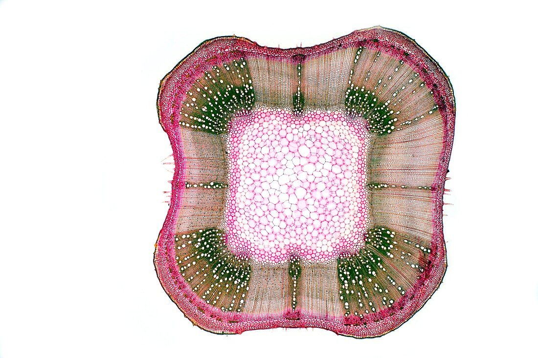
| Sage stem. Light micrograph of a section through a secondary stem of a scarlet sage (Salivia splendens) plant. The outer stem is covered with a thin epidermis and thick cuticle (red),but is thicker at the four corners forming cork (brown). It also contains stomata,and trichomes. Under the epidermis,at the corners,is a layer of flexible collenchyma (dark red) for support. Next is the cortex (red),with an inner pericycle of parenchyma tissue (brown). Next,the vascular bundles (green) consist or rings an outer layer of phloem (dark red) and inner xylem (green and brown) tissue,with cambium tissue (light red) in-between. At the centre of the stem is a pith (red) of parenchyma. Magnification: x5 when printed 10 centimetres wide | |
| Lizenzart: | Lizenzpflichtig |
| Credit: | Science Photo Library / Wheeler, Dr. Keith |
| Bildgröße: | 5125 px × 3417 px |
| Modell-Rechte: | nicht erforderlich |
| Restrictions: | - |
Preise für dieses Bild ab 15 €
Universitäten & Organisationen
(Informationsmaterial Digital, Informationsmaterial Print, Lehrmaterial Digital etc.)
ab 15 €
Redaktionell
(Bücher, Bücher: Sach- und Fachliteratur, Digitale Medien (redaktionell) etc.)
ab 30 €
Werbung
(Anzeigen, Aussenwerbung, Digitale Medien, Fernsehwerbung, Karten, Werbemittel, Zeitschriften etc.)
ab 55 €
Handelsprodukte
(bedruckte Textilie, Kalender, Postkarte, Grußkarte, Verpackung etc.)
ab 75 €
Pauschalpreise
Rechtepakete für die unbeschränkte Bildnutzung in Print oder Online
ab 495 €
Keywords
- Angiosperme,
- Angiospermen,
- ausgeschnitten,
- Ausschnitte,
- befleckt,
- Biologie,
- biologisch,
- Botanik,
- botanisch,
- Bündeln,
- Close-up,
- Cortex,
- Detail,
- Dikotyle,
- Epidermis,
- Gefäß,
- Gefäßband,
- Gefäße,
- Gewebe,
- Histologie,
- histologisch,
- Kambium,
- Kork,
- Kutikula,
- Lichtmikroskop,
- lichtmikroskopische Aufnahme,
- Mikroskopie,
- Parenchym,
- Pflanze,
- Pflanzen,
- Phloem,
- Salbei,
- Schicht,
- Schichten,
- Sektion,
- sektioniert,
- Stomata,
- Struktur,
- strukturell,
- Strukturen,
- Trichom,
- Trichome,
- Verfärbung,
- weißer Hintergrund,
- Xylem,
- Zellbilogie,
- Zelle,
- Zellen,
- Zytologie,
- Zytologisch