Lung tissue,fluorescence micrograph
Bildnummer 11611248
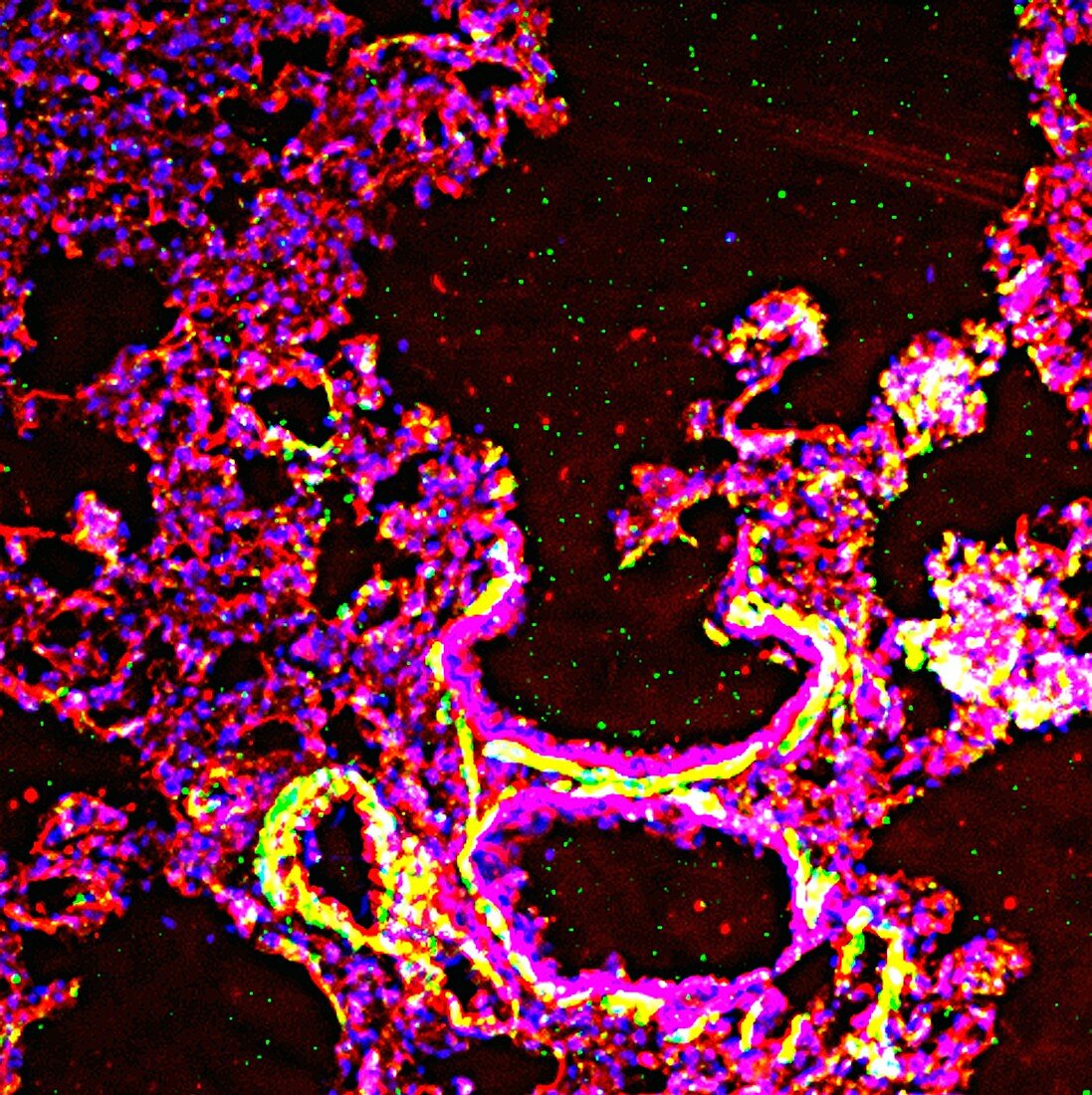
| Lung tissue. Fluorescence deconvolution micrograph of a section through lung tissue. This tissue sample shows the transition from a terminal bronchiole to a respiratory bronchiole. The walls of the airways are seen in cross-section at bottom. The lungs are where blood is oxygenated,with the oxygen replacing the waste product carbon dioxide that is produced during respiration in body cells. The bronchioles are branching airways that bring air from outside to the alveoli where gas exchange takes place. Cellular proteins are highlighted with fluorescent markers: g-actin (red),smooth muscle actin (green) and cell nuclei (blue) | |
| Lizenzart: | Lizenzpflichtig |
| Credit: | Science Photo Library / R. BICK, B. POINDEXTER, UT MEDICAL SCHOOL |
| Bildgröße: | 3005 px × 3012 px |
| Modell-Rechte: | nicht erforderlich |
| Eigentums-Rechte: | nicht erforderlich |
| Restrictions: | - |
Preise für dieses Bild ab 15 €
Universitäten & Organisationen
(Informationsmaterial Digital, Informationsmaterial Print, Lehrmaterial Digital etc.)
ab 15 €
Redaktionell
(Bücher, Bücher: Sach- und Fachliteratur, Digitale Medien (redaktionell) etc.)
ab 30 €
Werbung
(Anzeigen, Aussenwerbung, Digitale Medien, Fernsehwerbung, Karten, Werbemittel, Zeitschriften etc.)
ab 55 €
Handelsprodukte
(bedruckte Textilie, Kalender, Postkarte, Grußkarte, Verpackung etc.)
ab 75 €
Pauschalpreise
Rechtepakete für die unbeschränkte Bildnutzung in Print oder Online
ab 495 €
Keywords
- Aktin,
- Atemweg,
- Atemwege,
- Atmung,
- Atmungssystem,
- Biologie,
- biologisch,
- Bronchial,
- Bronchiolen,
- Farbstoff,
- Farbstoffe,
- Flecken,
- Fluoreszenz,
- g-Actin,
- gesund,
- Gewebe,
- Histologie,
- histologisch,
- Lichtmikroskop,
- lichtmikroskopische Aufnahme,
- Lunge,
- Lungen,
- Marker,
- menschlicher Körper,
- normal,
- Proteine,
- pulmonal,
- Querschnitt,
- respiratorische Bronchiole,
- Sektion,
- sektioniert,
- SMA,
- Verfärbung,
- Zellbilogie,
- Zelle,
- Zellen,
- Zellkern,
- zelluläres Protein,
- Zytologie,
- Zytologisch,
- Zytoskelett