Lung tissue,fluorescence micrograph
Bildnummer 11611246
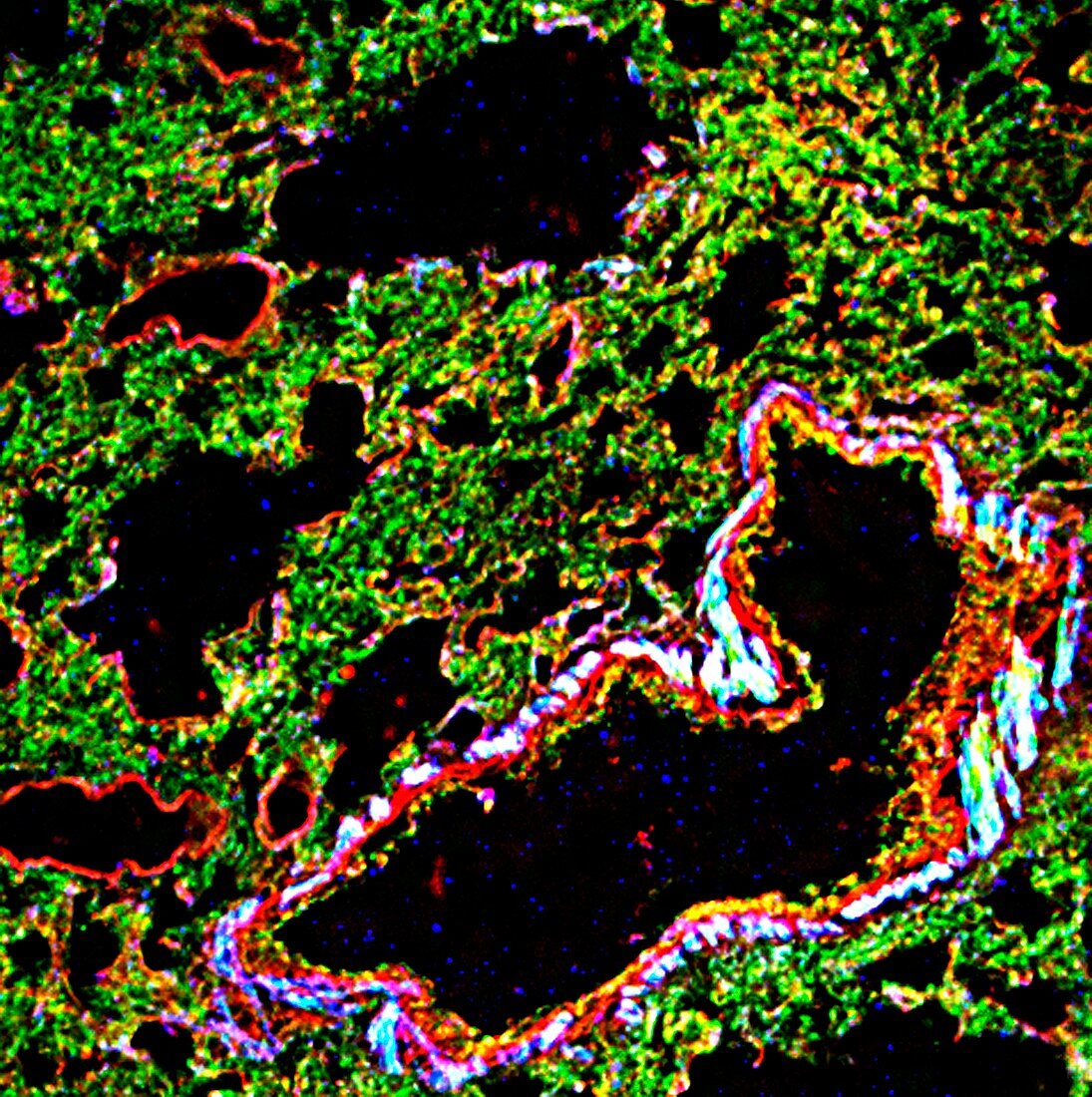
| Lung tissue. Fluorescence deconvolution micrograph of a section through lung tissue. This tissue sample shows a cross-section through an arteriole (small artery,black,lower right) in the lung at the point where it transitions to a thinner vessel (such as a capillary,black,upper left). The lungs are where blood is oxygenated,with the oxygen replacing the waste product carbon dioxide that is produced during respiration in body cells. Cellular proteins are highlighted with fluorescent markers: g-actin (red),f-actin (green) and smooth muscle actin (blue) | |
| Lizenzart: | Lizenzpflichtig |
| Credit: | Science Photo Library / R. BICK, B. POINDEXTER, UT MEDICAL SCHOOL |
| Bildgröße: | 2998 px × 3011 px |
| Modell-Rechte: | nicht erforderlich |
| Eigentums-Rechte: | nicht erforderlich |
| Restrictions: | - |
Preise für dieses Bild ab 15 €
Universitäten & Organisationen
(Informationsmaterial Digital, Informationsmaterial Print, Lehrmaterial Digital etc.)
ab 15 €
Redaktionell
(Bücher, Bücher: Sach- und Fachliteratur, Digitale Medien (redaktionell) etc.)
ab 30 €
Werbung
(Anzeigen, Aussenwerbung, Digitale Medien, Fernsehwerbung, Karten, Werbemittel, Zeitschriften etc.)
ab 55 €
Handelsprodukte
(bedruckte Textilie, Kalender, Postkarte, Grußkarte, Verpackung etc.)
ab 75 €
Pauschalpreise
Rechtepakete für die unbeschränkte Bildnutzung in Print oder Online
ab 495 €
Keywords
- Aktin,
- arteriell,
- Arteriole,
- Atmung,
- Atmungssystem,
- Biologie,
- biologisch,
- Blutgefäß,
- F-Actin,
- Farbstoff,
- Farbstoffe,
- Flecken,
- Fluoreszenz,
- g-Actin,
- Gefäße,
- gesund,
- Gewebe,
- Histologie,
- histologisch,
- kapillar,
- Kreislauf,
- Lichtmikroskop,
- lichtmikroskopische Aufnahme,
- Lumen,
- Lunge,
- Lungen,
- Marker,
- Mauer,
- menschlicher Körper,
- normal,
- Proteine,
- pulmonal,
- Querschnitt,
- Sektion,
- sektioniert,
- SMA,
- vaskulär,
- Verfärbung,
- Zellbilogie,
- Zelle,
- Zellen,
- zelluläres Protein,
- Zytologie,
- Zytologisch,
- Zytoskelett