Slipped disc,MRI scan
Bildnummer 11610416
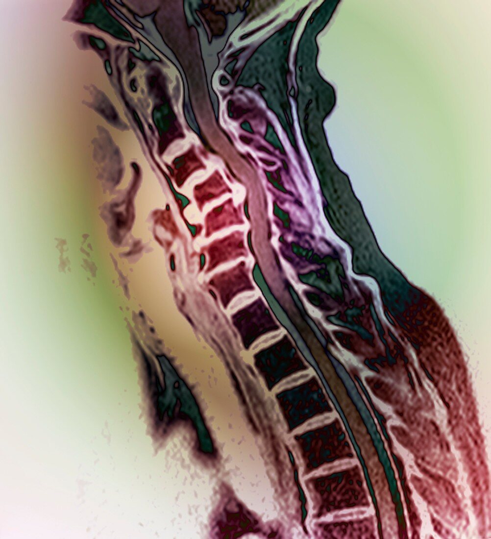
| Slipped disc,coloured magnetic resonance imaging (MRI) scan. Profile view of the spine of a 59-year-old patient with severe rheumatoid arthritis and a herniated (slipped) disc (red,upper centre-left). This condition occurs where one of the intervertebral discs (black bands) in the backbone ruptures. These discs are located between and cushion the vertebrae (squares). When a discs ruptures,the fluid and displaced tissue puts pressure on the nerves,causing pain. Here,one of the intervertebral discs in the neck (cervical region) has ruptured | |
| Lizenzart: | Lizenzpflichtig |
| Credit: | Science Photo Library / Zephyr |
| Bildgröße: | 4002 px × 4394 px |
| Modell-Rechte: | nicht erforderlich |
| Restrictions: | - |
Preise für dieses Bild ab 15 €
Universitäten & Organisationen
(Informationsmaterial Digital, Informationsmaterial Print, Lehrmaterial Digital etc.)
ab 15 €
Redaktionell
(Bücher, Bücher: Sach- und Fachliteratur, Digitale Medien (redaktionell) etc.)
ab 30 €
Werbung
(Anzeigen, Aussenwerbung, Digitale Medien, Fernsehwerbung, Karten, Werbemittel, Zeitschriften etc.)
ab 55 €
Handelsprodukte
(bedruckte Textilie, Kalender, Postkarte, Grußkarte, Verpackung etc.)
ab 75 €
Pauschalpreise
Rechtepakete für die unbeschränkte Bildnutzung in Print oder Online
ab 495 €
Keywords
- abnormal,
- arthritisch,
- Arthrologie,
- Bandscheibenvorfall,
- degenerativ,
- diagnostische Bildgebung,
- Erwachsene,
- farbig,
- Fünfziger Jahre,
- geduldig,
- gefärbt,
- Kondition,
- krank,
- Krankheit,
- Magnetresonanztomografie,
- Medizin,
- medizinisch,
- Mensch,
- Menschen,
- menschlicher Körper,
- MRI,
- Neurologie,
- neurologisch,
- Orthopädie,
- orthopädisch,
- Person,
- Profil,
- Radiographie,
- Radiologie,
- radiologisch,
- rheumatoide Arthritis,
- Rheumatologie,
- Rückgrat,
- Scan,
- Schmerz,
- schmerzhaft,
- Seitenansicht,
- Störung,
- ungesund,
- verletzt,
- Verletzung,
- Wirbel,
- Wirbelsäule,
- Wirbelsäulen-