Intestinal anatomy,artwork
Bildnummer 11609519
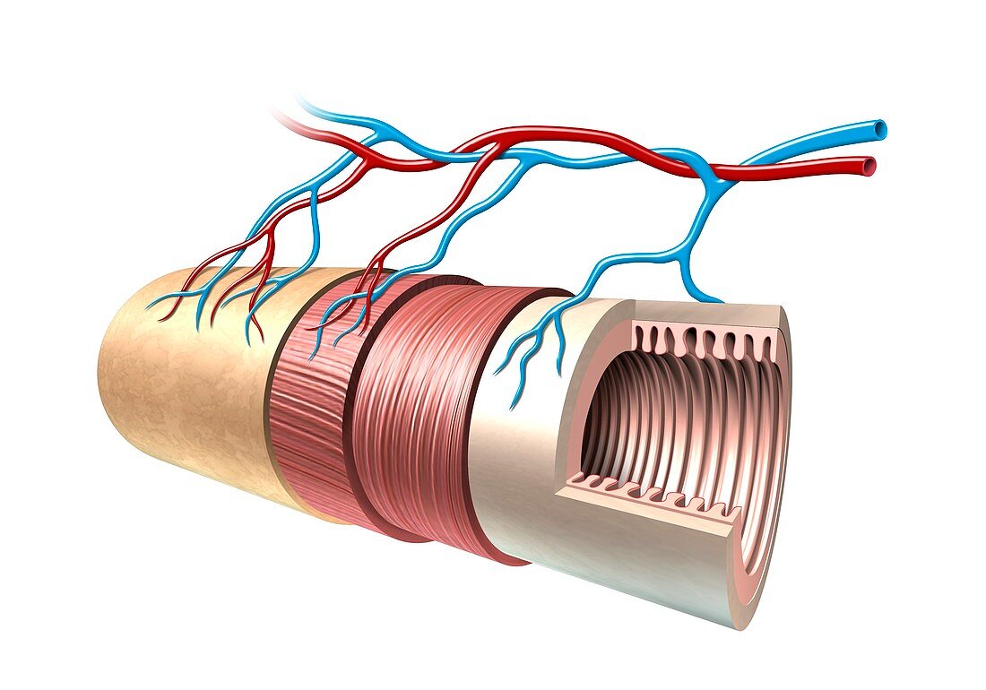
| Intestinal anatomy. Computer artwork showing the layers of the small intestine. The central space (lumen) is surrounded by the mucosa (beige,folded),which has numerous folds (villi). Surrounding this is the submucosa (beige) and then a layer of circular muscle (brown),followed by a layer of longitudinal muscle. The outer layer (beige,left) is the serous membrane (serosa). The arteries,which supply blood to the intestine,and veins that carry nutrients,absorbed during digestion,away from the intestine,can also be seen | |
| Lizenzart: | Lizenzpflichtig |
| Credit: | Science Photo Library / Dalhoff, Henning |
| Bildgröße: | 5007 px × 3503 px |
| Modell-Rechte: | nicht erforderlich |
| Restrictions: | - |
Preise für dieses Bild ab 15 €
Universitäten & Organisationen
(Informationsmaterial Digital, Informationsmaterial Print, Lehrmaterial Digital etc.)
ab 15 €
Redaktionell
(Bücher, Bücher: Sach- und Fachliteratur, Digitale Medien (redaktionell) etc.)
ab 30 €
Werbung
(Anzeigen, Aussenwerbung, Digitale Medien, Fernsehwerbung, Karten, Werbemittel, Zeitschriften etc.)
ab 55 €
Handelsprodukte
(bedruckte Textilie, Kalender, Postkarte, Grußkarte, Verpackung etc.)
ab 75 €
Pauschalpreise
Rechtepakete für die unbeschränkte Bildnutzung in Print oder Online
ab 495 €
Keywords
- Anatomie,
- anatomisch,
- Arterie,
- Arterien,
- ausgeschnitten,
- Ausschnitte,
- Biologie,
- biologisch,
- Blutversorgung,
- Darm,
- Darm-,
- Dünndarm,
- Falten,
- Gastroenterologie,
- gastrointestinal,
- Gefäßsystem,
- gesund,
- GI tract,
- Illustration,
- Kunstwerk,
- Lumen,
- Mensch,
- menschlicher Körper,
- Muskeln,
- normal,
- Physiologie,
- physiologisch,
- Schicht,
- Schichten,
- Schleimhaut,
- serosa,
- Struktur,
- Strukturen,
- Submukosa,
- System,
- Vene,
- Venen,
- weißer Hintergrund,
- Zotte,
- Zotten