Nasal mucosa,artwork
Bildnummer 11604347
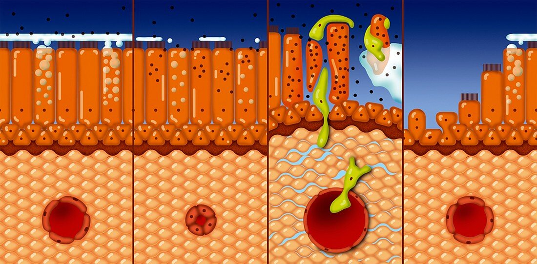
| Nasal mucosa,cross-section. Artwork of a sequence (left to right) showing mucus production in response to infection and inflammation. The surface is at top,with epithelial cells normally covered by a thin,clear layer of mucus that helps to filter incoming air. When infection occurs,this epithelium starts to break down (second image). By the third image,thick mucus (green) is being produced by glands in the underlying layer (lamina propria). One of these glands is shown here. Swelling and inflammation is also seen. By the fourth image,the swelling has subsided,and the epithelium is starting to regenerate | |
| Lizenzart: | Lizenzpflichtig |
| Credit: | Science Photo Library / Art for Science |
| Bildgröße: | 5991 px × 2948 px |
| Modell-Rechte: | nicht erforderlich |
| Eigentums-Rechte: | nicht erforderlich |
| Restrictions: | - |
Preise für dieses Bild ab 15 €
Universitäten & Organisationen
(Informationsmaterial Digital, Informationsmaterial Print, Lehrmaterial Digital etc.)
ab 15 €
Redaktionell
(Bücher, Bücher: Sach- und Fachliteratur, Digitale Medien (redaktionell) etc.)
ab 30 €
Werbung
(Anzeigen, Aussenwerbung, Digitale Medien, Fernsehwerbung, Karten, Werbemittel, Zeitschriften etc.)
ab 55 €
Handelsprodukte
(bedruckte Textilie, Kalender, Postkarte, Grußkarte, Verpackung etc.)
ab 75 €
Pauschalpreise
Rechtepakete für die unbeschränkte Bildnutzung in Print oder Online
ab 495 €
Keywords
- Biologie,
- biologisch,
- Drüse,
- Drüsen,
- entzündet,
- Entzündung,
- Epithel,
- epithelial,
- geschwollen,
- Gewebe,
- Illustration,
- Kalt,
- Kunstwerk,
- Medizin,
- medizinisch,
- menschlicher Körper,
- Nasal-,
- Nase,
- Physiologie,
- physiologisch,
- Querschnitt,
- Regeneration,
- Reihenfolge,
- Schleim,
- Schleimhaut,
- Sektion,
- sektioniert,
- Serie,
- Submukosa,
- Zellbilogie,
- Zelle,
- zerstörend