Intestinal villus cell loss,SEM
Bildnummer 11602616
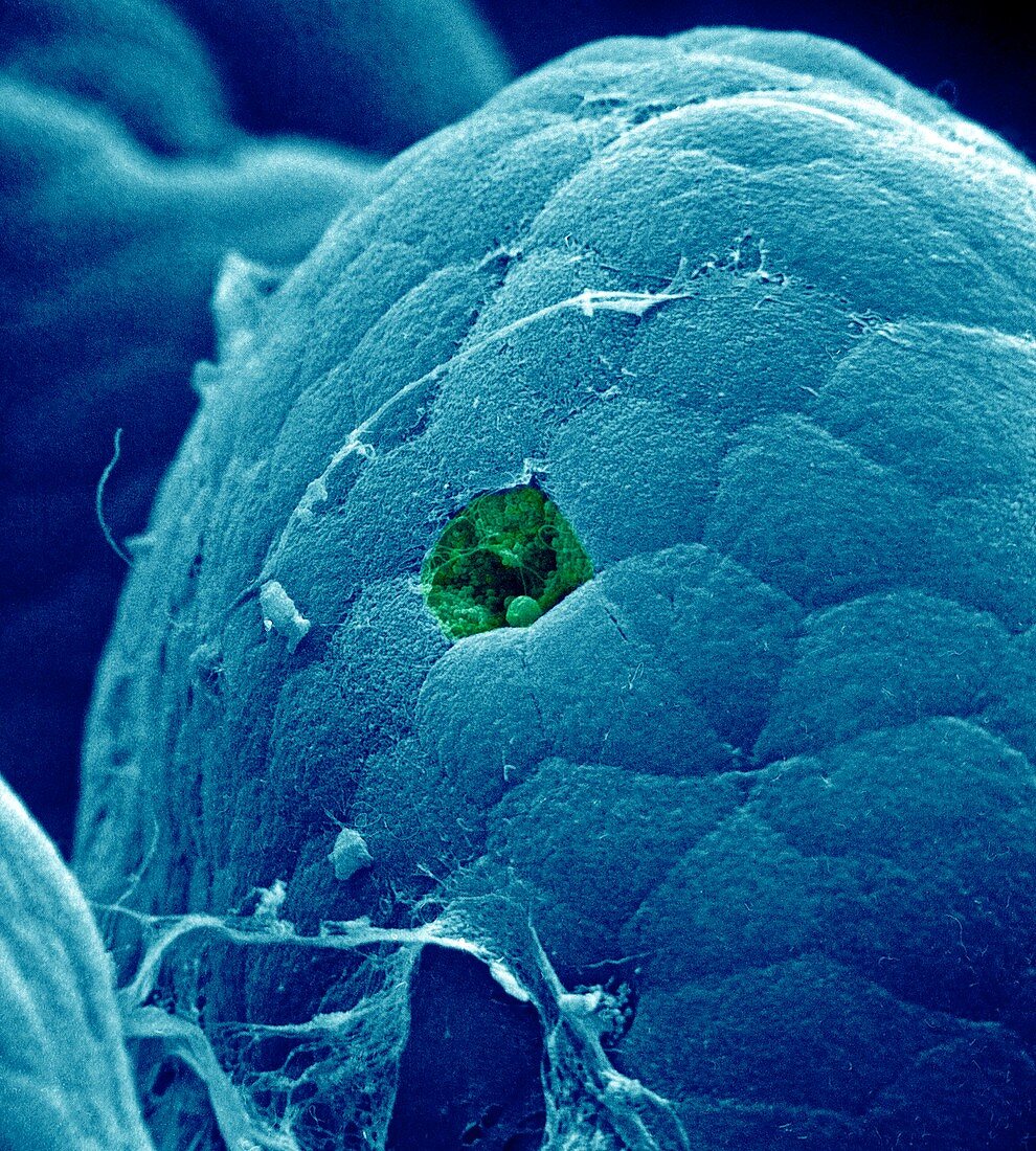
| Intestinal villus cell loss,coloured scanning electron micrograph (SEM). Close-up of the tip of a villus in the small intestine that has shed a cell from its tip as part of the normal cell replacement cycle,leaving a void and cell debris (green) that will swiftly be sealed to prevent infection. The villi are the finger-like projections that line the surface of the small intestine and absorb nutrients from digested food. The surface of each villus is covered by hundreds of cells called enterocytes,which migrate from the base (where they are formed) to the tip,being shed at the rate of around one a minute. Magnification: x1200 when printed 10 centimetres wide | |
| Lizenzart: | Lizenzpflichtig |
| Credit: | Science Photo Library |
| Bildgröße: | 2822 px × 3134 px |
| Modell-Rechte: | nicht erforderlich |
| Eigentums-Rechte: | nicht erforderlich |
| Restrictions: | - |
Preise für dieses Bild ab 15 €
Universitäten & Organisationen
(Informationsmaterial Digital, Informationsmaterial Print, Lehrmaterial Digital etc.)
ab 15 €
Redaktionell
(Bücher, Bücher: Sach- und Fachliteratur, Digitale Medien (redaktionell) etc.)
ab 30 €
Werbung
(Anzeigen, Aussenwerbung, Digitale Medien, Fernsehwerbung, Karten, Werbemittel, Zeitschriften etc.)
ab 55 €
Handelsprodukte
(bedruckte Textilie, Kalender, Postkarte, Grußkarte, Verpackung etc.)
ab 75 €
Pauschalpreise
Rechtepakete für die unbeschränkte Bildnutzung in Print oder Online
ab 495 €
Keywords
- Anatomie,
- anatomisch,
- Apoptose,
- Biologie,
- Close-up,
- Darm,
- Darm-,
- Detail,
- Dünndarm,
- eingefärbt,
- Enterozyten,
- Epithel,
- epithelial,
- farbig,
- Gedärme,
- gefärbt,
- gesund,
- Gewebe,
- Histologie,
- histologisch,
- Lumen,
- Magen-Darm-Trakt,
- menschlicher Körper,
- normal,
- Physiologie,
- physiologisch,
- Rasterelektronenmikroskop,
- rasterelektronenmikroskopische Aufnahme,
- Ratschlag,
- REM,
- Schuppen,
- Verdauungskanal,
- Verdauungssystem,
- Zellbilogie,
- Zelle,
- Zellen,
- Zellersatz,
- Zotte