Liana stem,light micrograph
Bildnummer 11593536
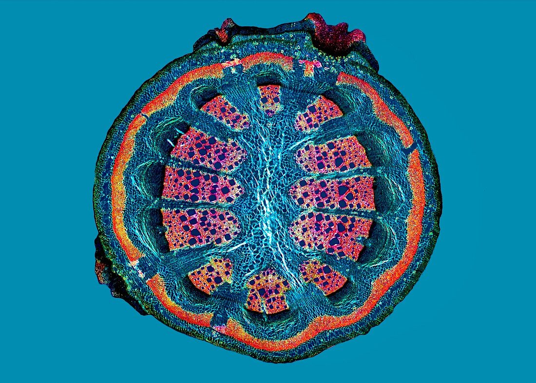
| Liana stem. Polarised light micrograph,of a cross-section through the stem of a climbing liana (Aristolochia tormentosa). Two thin outer tissue layers contain collenchyma (green) and parenchyma (blue). This is followed by three thin vascular layers: the outer fibre bundles (orange); phloem (deep blue); and cambium (black). A larger vascular layer is the xylem (pink),with medullary rays (blue) passing through it to the central pith (blue) of parenchyma cells. Magnification: x44 when printed at 10 centimetres across | |
| Lizenzart: | Lizenzpflichtig |
| Credit: | Science Photo Library / Wheeler, Dr. Keith |
| Bildgröße: | 4961 px × 3543 px |
| Modell-Rechte: | nicht erforderlich |
| Eigentums-Rechte: | nicht erforderlich |
| Restrictions: | - |
Preise für dieses Bild ab 15 €
Universitäten & Organisationen
(Informationsmaterial Digital, Informationsmaterial Print, Lehrmaterial Digital etc.)
ab 15 €
Redaktionell
(Bücher, Bücher: Sach- und Fachliteratur, Digitale Medien (redaktionell) etc.)
ab 30 €
Werbung
(Anzeigen, Aussenwerbung, Digitale Medien, Fernsehwerbung, Karten, Werbemittel, Zeitschriften etc.)
ab 55 €
Handelsprodukte
(bedruckte Textilie, Kalender, Postkarte, Grußkarte, Verpackung etc.)
ab 75 €
Pauschalpreise
Rechtepakete für die unbeschränkte Bildnutzung in Print oder Online
ab 495 €
Keywords
- ausgeschnitten,
- Ausschnitte,
- Biologie,
- biologisch,
- Botanik,
- botanisch,
- diagonal,
- Flora,
- Gefäßband,
- Gewebe,
- Kreis,
- kreisförmig,
- Lichtmikroskop,
- lichtmikroskopische Aufnahme,
- Natur,
- Pflanze,
- Phloem,
- plm,
- polarisiert,
- Querschnitt,
- rund,
- Sektion,
- sektioniert,
- Stengel,
- Wassertransport,
- Weinrebe,
- Xylem,
- Zelle,
- Zellen,
- zellular