Secondary liver cancer,3D angiogram
Bildnummer 11589633
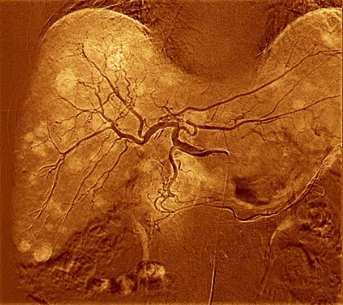
| Secondary cancer in the liver. Coloured 3D angiogram (blood vessel X-ray) of the blood vessels in the liver of a 67-year-old patient with secondary (metastatic) cancer in the liver. Lesions (white patches) show where the cancers have spread to the liver from a primary cancer elsewhere. This image was obtained as part of preparations for intra-hepatic chemotherapy by injections into the blood vessels. This image was produced by Digital Subtraction Angiography (DSA),using a contrast medium to highlight the blood vessels | |
| Lizenzart: | Lizenzpflichtig |
| Credit: | Science Photo Library / Zephyr |
| Bildgröße: | 3147 px × 2803 px |
| Modell-Rechte: | nicht erforderlich |
| Eigentums-Rechte: | nicht erforderlich |
| Restrictions: | - |
Preise für dieses Bild ab 15 €
Universitäten & Organisationen
(Informationsmaterial Digital, Informationsmaterial Print, Lehrmaterial Digital etc.)
ab 15 €
Redaktionell
(Bücher, Bücher: Sach- und Fachliteratur, Digitale Medien (redaktionell) etc.)
ab 30 €
Werbung
(Anzeigen, Aussenwerbung, Digitale Medien, Fernsehwerbung, Karten, Werbemittel, Zeitschriften etc.)
ab 55 €
Handelsprodukte
(bedruckte Textilie, Kalender, Postkarte, Grußkarte, Verpackung etc.)
ab 75 €
Pauschalpreise
Rechtepakete für die unbeschränkte Bildnutzung in Print oder Online
ab 495 €
Keywords
- 3-d,
- 3D,
- 60er Jahre,
- abnormal,
- Angiografie,
- Angiogramm,
- anterior,
- Arterie,
- Arterien,
- Ausbreitung,
- Behandlung,
- Blutgefäß,
- Blutgefäße,
- Chemotherapie,
- Diagnose,
- digitale Subtraktionsangiographie,
- Dreidimensional,
- dsa,
- Einfarbig,
- eingefärbt,
- Erwachsene,
- farbig,
- Fehlfarbe,
- Frontal,
- geduldig,
- gefärbt,
- hepatisch,
- Hepatologie,
- Kondition,
- Kontrastmittel,
- Krankheit,
- Krebs,
- krebsartig,
- Kreislauf,
- Leber,
- maligne,
- Medizin,
- medizinisch,
- menschlicher Körper,
- Metastase,
- mittleren Alters,
- Onkologie,
- onkologisch,
- Orange,
- Radiographie,
- Röntgen,
- Röntgengerät,
- sechziger Jahre,
- sekundär,
- Störung,
- ungesund,
- vaskulär,
- Vene,
- Venen