Keratoacanthoma,light micrograph
Bildnummer 11584769
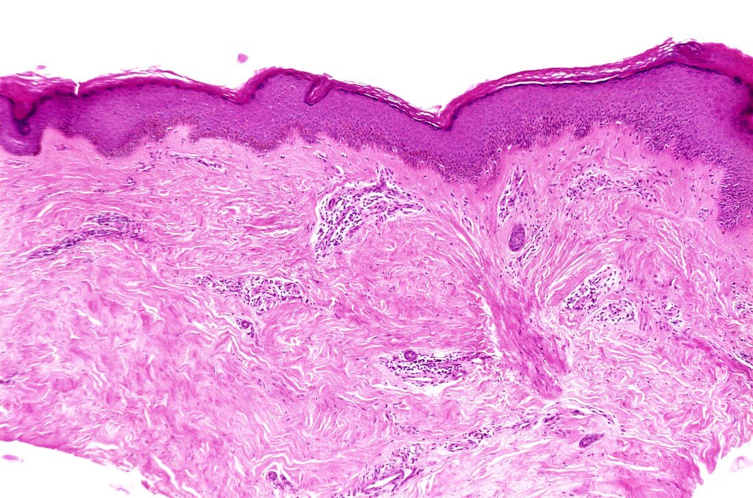
| Keratoacanthoma. Light micrograph of a section through a keratoacanthoma nodule. The surface layers are across top,including the hard keratin layer. Below this there are patches of inflammation and white blood cells with stained nuclei (dark spots). The cause of a keratoacanthoma lesion is unknown but it has many features of a viral condition,and consists of a localised proliferation of squamous cells that forms a cratered nodule. The nodule grows over several weeks before gradually disappearing. However,the unsightly nodule is often surgically removed. The cause of keratoacanthoma is unknown,although exposure to sunlight appears to be a factor | |
| Lizenzart: | Lizenzpflichtig |
| Credit: | Science Photo Library / Biophoto Associates |
| Bildgröße: | 5179 px × 3425 px |
| Modell-Rechte: | nicht erforderlich |
| Eigentums-Rechte: | nicht erforderlich |
| Restrictions: |
|
Preise für dieses Bild ab 15 €
Universitäten & Organisationen
(Informationsmaterial Digital, Informationsmaterial Print, Lehrmaterial Digital etc.)
ab 15 €
Redaktionell
(Bücher, Bücher: Sach- und Fachliteratur, Digitale Medien (redaktionell) etc.)
ab 30 €
Werbung
(Anzeigen, Aussenwerbung, Digitale Medien, Fernsehwerbung, Karten, Werbemittel, Zeitschriften etc.)
ab 55 €
Handelsprodukte
(bedruckte Textilie, Kalender, Postkarte, Grußkarte, Verpackung etc.)
ab 75 €
Pauschalpreise
Rechtepakete für die unbeschränkte Bildnutzung in Print oder Online
ab 495 €
Keywords
- abnormal,
- chronische Entzündung,
- dermal,
- Dermatologie,
- dermatologisch,
- Diagnose,
- entzündet,
- Entzündung,
- Entzündungsreaktion,
- epidermal,
- Epidermis,
- Haut,
- Histologie,
- histologisch,
- Histopathologie,
- histopathologisch,
- Keratin,
- Keratoakanthom,
- Kondition,
- Krankheit,
- Lichtmikroskop,
- lichtmikroskopische Aufnahme,
- Masse,
- Medizin,
- medizinisch,
- menschlicher Körper,
- Oberfläche,
- Pathologie,
- Schicht,
- Schichten,
- Störung,
- ungesund,
- Wachstum,
- weißes Blutkörperchen,
- Wunde,
- Zellen