Oesophagus lining
Bildnummer 11571587
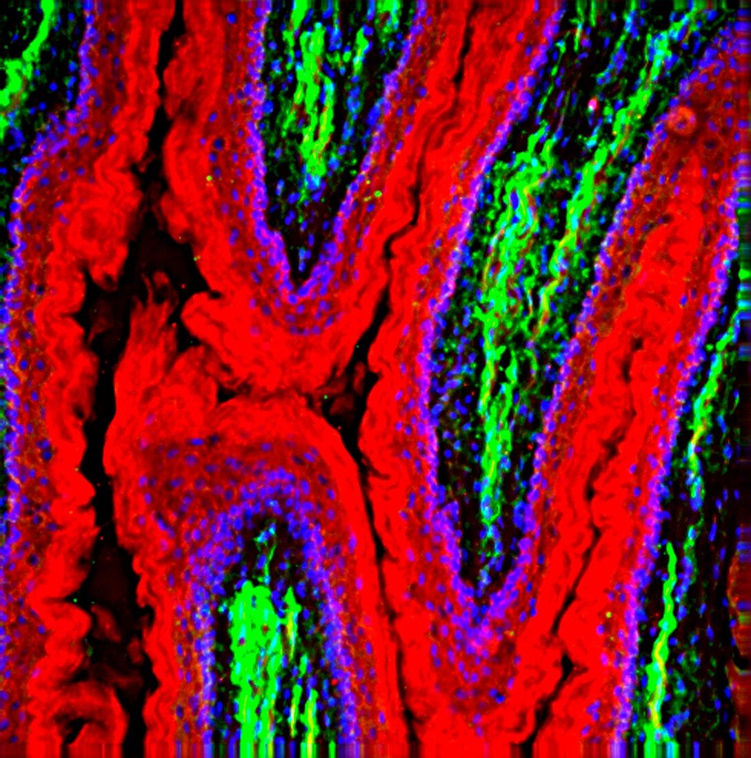
| Oesophagus lining. Fluorescence deconvolution micrograph of a section through a healthy oesophagus (gullet),showing the muscle lining (g-actin,red). Green is the f-actin of muscle in the walls,and blue is cell nuclei. Magnification x40 when printed 10 centimetres wide | |
| Lizenzart: | Lizenzpflichtig |
| Credit: | Science Photo Library / R. BICK, B. POINDEXTER, UT MEDICAL SCHOOL |
| Bildgröße: | 2946 px × 2976 px |
| Modell-Rechte: | nicht erforderlich |
| Eigentums-Rechte: | nicht erforderlich |
| Restrictions: | - |
Preise für dieses Bild ab 15 €
Universitäten & Organisationen
(Informationsmaterial Digital, Informationsmaterial Print, Lehrmaterial Digital etc.)
ab 15 €
Redaktionell
(Bücher, Bücher: Sach- und Fachliteratur, Digitale Medien (redaktionell) etc.)
ab 30 €
Werbung
(Anzeigen, Aussenwerbung, Digitale Medien, Fernsehwerbung, Karten, Werbemittel, Zeitschriften etc.)
ab 55 €
Handelsprodukte
(bedruckte Textilie, Kalender, Postkarte, Grußkarte, Verpackung etc.)
ab 75 €
Pauschalpreise
Rechtepakete für die unbeschränkte Bildnutzung in Print oder Online
ab 495 €
Keywords
- Aktin,
- Anatomie,
- anatomisch,
- Atomkern,
- Biologie,
- biologisch,
- Eiweiß,
- Fluoreszenz,
- g-Actin,
- gastrointestinal,
- gesund,
- Gewebe,
- Histologie,
- histologisch,
- histopathologisch,
- Kerne,
- Lichtmikroskop,
- lichtmikroskopische Aufnahme,
- Mauer,
- Medizin,
- medizinisch,
- Mensch,
- menschlicher Körper,
- Mikrofotografie,
- Muskeln,
- Muskulös,
- normal,
- Oesophagus,
- Person,
- Sektion,
- sektioniert,
- Speiseröhre,
- Struktur,
- Zelle,
- Zellen