Keratoacanthoma,light micrograph
Bildnummer 11569117
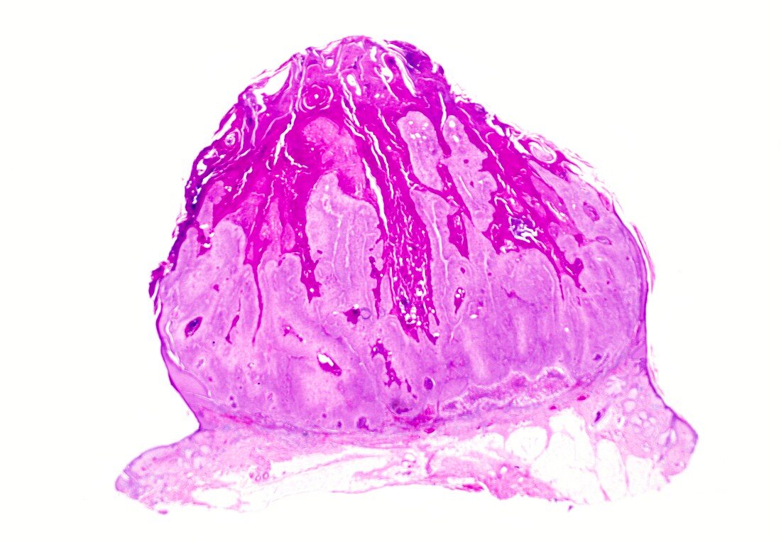
| Keratoacanthoma. Light micrograph of a section through a keratoacanthoma nodule. The cause of these lesions is unknown but they have many features of a viral condition. A localised proliferation of squamous cells (light pink) have produced the nodule,with a central crater containing a mass of keratin (dark pink). The peripheral epidermis is normal and the dermis shows a chronic,heavy infiltration of inflammatory cells | |
| Lizenzart: | Lizenzpflichtig |
| Credit: | Science Photo Library / Biophoto Associates |
| Bildgröße: | 5079 px × 3522 px |
| Modell-Rechte: | nicht erforderlich |
| Eigentums-Rechte: | nicht erforderlich |
| Restrictions: |
|
Preise für dieses Bild ab 15 €
Universitäten & Organisationen
(Informationsmaterial Digital, Informationsmaterial Print, Lehrmaterial Digital etc.)
ab 15 €
Redaktionell
(Bücher, Bücher: Sach- und Fachliteratur, Digitale Medien (redaktionell) etc.)
ab 30 €
Werbung
(Anzeigen, Aussenwerbung, Digitale Medien, Fernsehwerbung, Karten, Werbemittel, Zeitschriften etc.)
ab 55 €
Handelsprodukte
(bedruckte Textilie, Kalender, Postkarte, Grußkarte, Verpackung etc.)
ab 75 €
Pauschalpreise
Rechtepakete für die unbeschränkte Bildnutzung in Print oder Online
ab 495 €
Keywords
- abnormal,
- Anatomie,
- anatomisch,
- Biologie,
- biologisch,
- chronische Entzündung,
- dermal,
- Dermatologie,
- dermatologisch,
- entzündet,
- epidermal,
- Epidermis,
- Haut,
- Histologie,
- histologisch,
- Histopathologie,
- histopathologisch,
- Keratin,
- Keratoakanthom,
- Kondition,
- Krankheit,
- Lichtmikroskop,
- lichtmikroskopische Aufnahme,
- Masse,
- Medizin,
- medizinisch,
- Pathologie,
- rosa,
- Schicht,
- Schichten,
- Störung,
- ungesund,
- weißer Hintergrund,
- Wunde,
- Zellen