Dislocated hip replacement,X-ray
Bildnummer 11566359
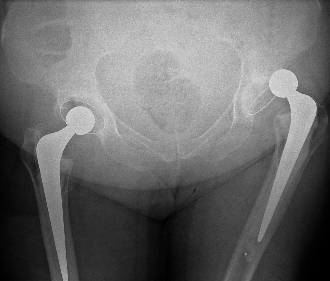
| Dislocated hip replacement. Frontal X-ray of the pelvis of a patient with bi-lateral (both sides) total hip replacements. The right joint replacement (right) has become dislocated. The metal ball-and-shaft implant (solid white) in the thigh bone (femur) has pushed out of the artificial socket (cup-shaped,centre right) with which it should articulate and form a joint in the pelvis | |
| Lizenzart: | Lizenzpflichtig |
| Credit: | Science Photo Library / Du Cane Medical Imaging |
| Bildgröße: | 4571 px × 3897 px |
| Modell-Rechte: | nicht erforderlich |
| Eigentums-Rechte: | nicht erforderlich |
| Restrictions: | - |
Preise für dieses Bild ab 15 €
Universitäten & Organisationen
(Informationsmaterial Digital, Informationsmaterial Print, Lehrmaterial Digital etc.)
ab 15 €
Redaktionell
(Bücher, Bücher: Sach- und Fachliteratur, Digitale Medien (redaktionell) etc.)
ab 30 €
Werbung
(Anzeigen, Aussenwerbung, Digitale Medien, Fernsehwerbung, Karten, Werbemittel, Zeitschriften etc.)
ab 55 €
Handelsprodukte
(bedruckte Textilie, Kalender, Postkarte, Grußkarte, Verpackung etc.)
ab 75 €
Pauschalpreise
Rechtepakete für die unbeschränkte Bildnutzung in Print oder Online
ab 495 €
Keywords
- abnormal,
- Arthrologie,
- Becken,
- behandelt,
- Behandlung,
- Einfarbig,
- geduldig,
- Gelenke,
- Gesundheitswesen,
- Knochen,
- Kondition,
- Krankheit,
- künstlich,
- Medizin,
- medizinisch,
- Mensch,
- Menschen,
- menschlicher Körper,
- Metall,
- Orthopädie,
- orthopädisch,
- Osteologie,
- pelvin,
- Person,
- Prothese,
- Radiographie,
- Radiologie,
- Rheumatologie,
- Röntgen,
- Röntgengerät,
- Schwarz und weiß,
- Störung,
- ungesund,
- Vorderansicht