Elm stem,light micrograph
Bildnummer 11565158
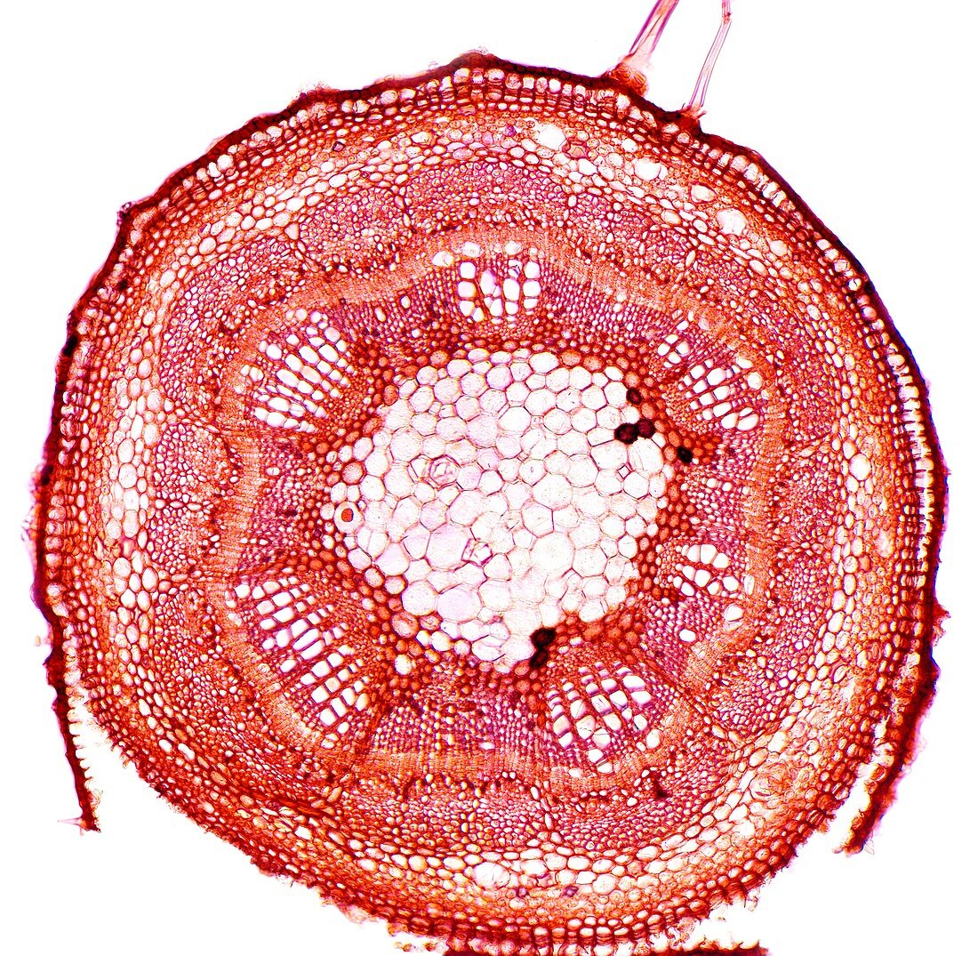
| Elm stem. Light micrograph of a transverse section through the one-year-old woody stem of an elm (Ulmus procera) tree. The outer layer (epidermis,dark ring) is being sloughed (pushed/broken) off by developing underlying cork (red-brown),underneath which is the cortex made of collenchyma (orange-red). The lower cortex has bands of phloem fibres (red) and a layer of phloem sieve tubes (solid red with cavities). Under the phloem is a ring of cambium (light-red circle) and under this is a thick layer of wood (xylem,purple-red) with six vascular bundles. The solid pith (pink,centre) is composed of parenchyma cells. Magnification: x46 when printed 10 centimetres wide | |
| Lizenzart: | Lizenzpflichtig |
| Credit: | Science Photo Library / Wheeler, Dr. Keith |
| Bildgröße: | 5362 px × 5241 px |
| Modell-Rechte: | nicht erforderlich |
| Eigentums-Rechte: | nicht erforderlich |
| Restrictions: | - |
Preise für dieses Bild ab 15 €
Universitäten & Organisationen
(Informationsmaterial Digital, Informationsmaterial Print, Lehrmaterial Digital etc.)
ab 15 €
Redaktionell
(Bücher, Bücher: Sach- und Fachliteratur, Digitale Medien (redaktionell) etc.)
ab 30 €
Werbung
(Anzeigen, Aussenwerbung, Digitale Medien, Fernsehwerbung, Karten, Werbemittel, Zeitschriften etc.)
ab 55 €
Handelsprodukte
(bedruckte Textilie, Kalender, Postkarte, Grußkarte, Verpackung etc.)
ab 75 €
Pauschalpreise
Rechtepakete für die unbeschränkte Bildnutzung in Print oder Online
ab 495 €
Keywords
- Angiosperme,
- Angiospermen,
- Biologie,
- biologisch,
- Botanik,
- botanisch,
- Cortex,
- Fasern,
- Flora,
- Gefäße,
- Kambium,
- Kork,
- Lichtmikroskop,
- lichtmikroskopische Aufnahme,
- Natur,
- Parenchym,
- Pflanze,
- primäre Phloemfaser,
- Ringe,
- Sektion,
- sektioniert,
- Stengel,
- unterstützend,
- Unterstützung,
- weißer Hintergrund,
- Xylemgefäß,
- Zellbilogie,
- Zelle,
- Zellen,
- Zytologie