Cedar tree stem,light micrograph
Bildnummer 11564937
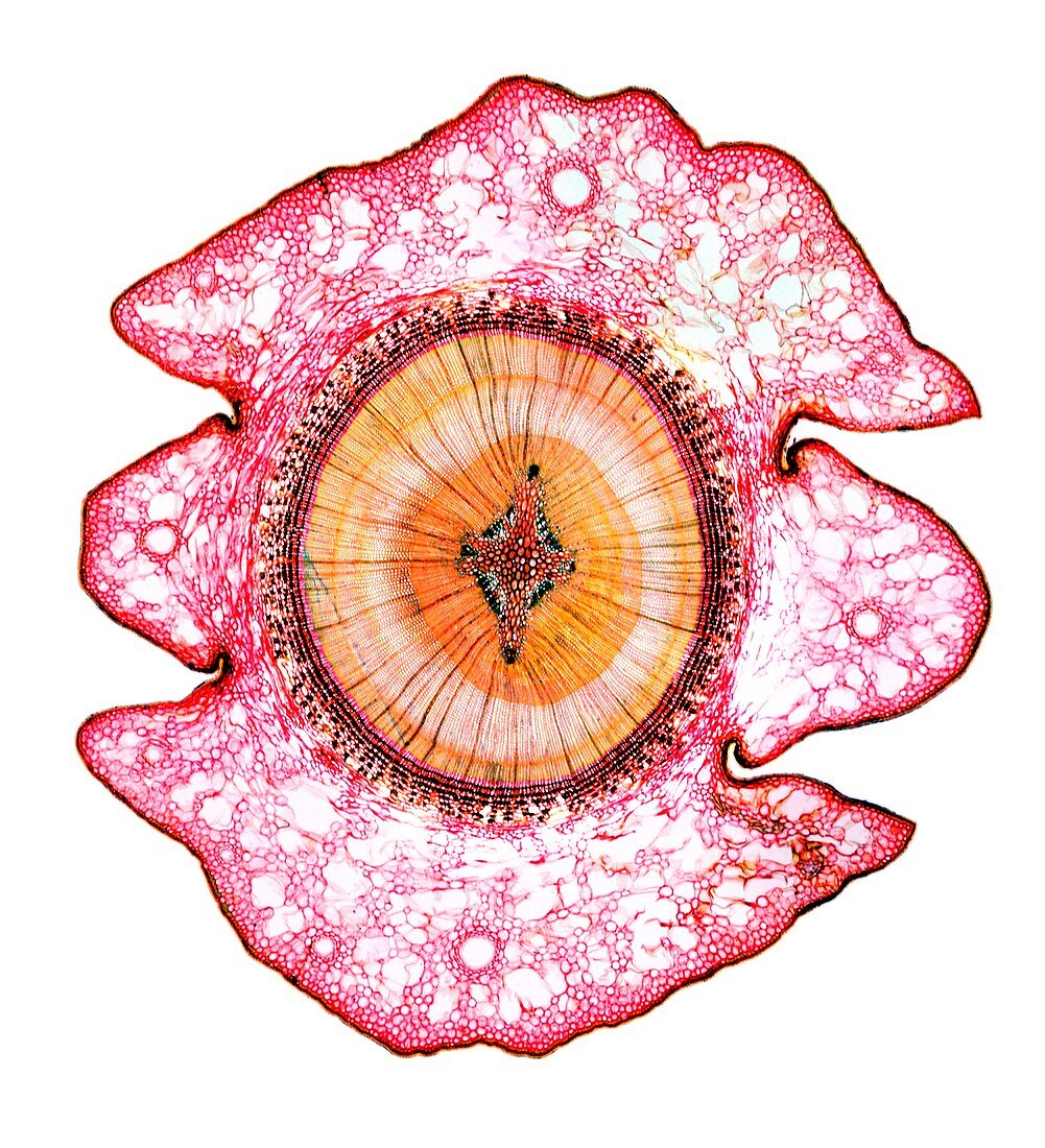
| Cedar tree stem. Light micrograph of a transverse section through a stem of a cedar tree (Thujopsis dolobrata). The four ridges on the outer surface are microphyllous leaves,which surround the stem with their inner surfaces fused to the stem cortex. On the outside of the vascular cylinder (centre,round) is the secondary phloem consisting of sieve cells (pink bands) and phloem fibres (black bands). The inner xylem (orange) is made up of thick walled tracheid cells. Radial lines (dark orange) are the single celled rays composed of tracheid cells. In-between the phloem and xylem is the cambium (red band). Magnification: x14 when printed 10 centimetres wide | |
| Lizenzart: | Lizenzpflichtig |
| Credit: | Science Photo Library / Wheeler, Dr. Keith |
| Bildgröße: | 4080 px × 4483 px |
| Modell-Rechte: | nicht erforderlich |
| Eigentums-Rechte: | nicht erforderlich |
| Restrictions: | - |
Preise für dieses Bild ab 15 €
Universitäten & Organisationen
(Informationsmaterial Digital, Informationsmaterial Print, Lehrmaterial Digital etc.)
ab 15 €
Redaktionell
(Bücher, Bücher: Sach- und Fachliteratur, Digitale Medien (redaktionell) etc.)
ab 30 €
Werbung
(Anzeigen, Aussenwerbung, Digitale Medien, Fernsehwerbung, Karten, Werbemittel, Zeitschriften etc.)
ab 55 €
Handelsprodukte
(bedruckte Textilie, Kalender, Postkarte, Grußkarte, Verpackung etc.)
ab 75 €
Pauschalpreise
Rechtepakete für die unbeschränkte Bildnutzung in Print oder Online
ab 495 €
Keywords
- ausgeschnitten,
- Ausschnitte,
- Baum,
- Biologie,
- biologisch,
- Blatt,
- Botanik,
- botanisch,
- Epidermis,
- Flora,
- Gefäßband,
- gymnospermen,
- Jung,
- Kambium,
- Kutikula,
- Lichtmikroskop,
- lichtmikroskopische Aufnahme,
- Nacktsamer,
- Nadelbaum,
- Natur,
- Pflanze,
- Phloem,
- Sektion,
- sektioniert,
- Stengel,
- unterstützend,
- Unterstützung,
- weißer Hintergrund,
- Xylem,
- zapfentragend,
- Zeder,
- Zellbilogie,
- Zelle,
- Zellen,
- Zytologie