Tea leaf,light micrograph
Bildnummer 11564427
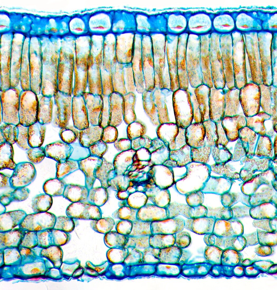
| Tea leaf. Light micrograph of a cross-section through a tea (Camellia sinensis) leaf. The upper and lower epidermis on the surfaces of the leaf are blue. Under the upper epidermis are palisade cells (brown),which contain chloroplasts,the site of photosynthesis. Beneath this a spongy mesophyll layer with large spaces between the cells. At bottom left,a stoma (pore) is seen. Stomata allow gases and water to enter and leave the plant. Magnification: x230 when printed 10 centimetres wide | |
| Lizenzart: | Lizenzpflichtig |
| Credit: | Science Photo Library / Wheeler, Dr. Keith |
| Bildgröße: | 5171 px × 5417 px |
| Modell-Rechte: | nicht erforderlich |
| Eigentums-Rechte: | nicht erforderlich |
| Restrictions: | - |
Preise für dieses Bild ab 15 €
Universitäten & Organisationen
(Informationsmaterial Digital, Informationsmaterial Print, Lehrmaterial Digital etc.)
ab 15 €
Redaktionell
(Bücher, Bücher: Sach- und Fachliteratur, Digitale Medien (redaktionell) etc.)
ab 30 €
Werbung
(Anzeigen, Aussenwerbung, Digitale Medien, Fernsehwerbung, Karten, Werbemittel, Zeitschriften etc.)
ab 55 €
Handelsprodukte
(bedruckte Textilie, Kalender, Postkarte, Grußkarte, Verpackung etc.)
ab 75 €
Pauschalpreise
Rechtepakete für die unbeschränkte Bildnutzung in Print oder Online
ab 495 €