'Herniated spinal disc,MRI scan'
Bildnummer 11560741
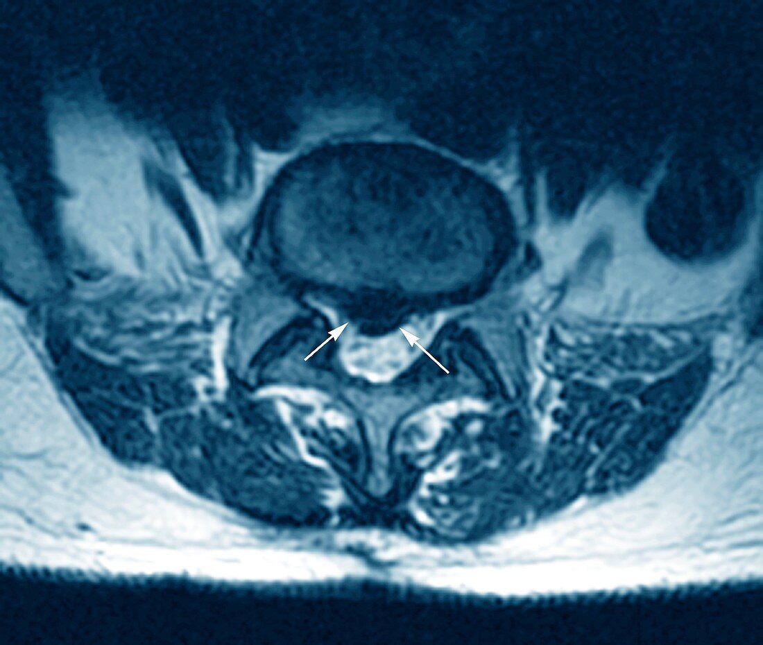
| Magnetic resonance imaging (MRI) scan of an axial (horizontal) view of the spine of a patient aged 35,showing a herniated disc at upper centre (rounded) which has ruptured (arrows) causing it to press on the spinal cord below (white). This disc is between lumbar vertebra (L5) and sacral vertebra (S1) in the lower back. Surgery was required to replace this damaged disc with an artificial (prosthetic) one | |
| Lizenzart: | Lizenzpflichtig |
| Credit: | Science Photo Library / Zephyr |
| Bildgröße: | 4569 px × 3862 px |
| Modell-Rechte: | nicht erforderlich |
| Eigentums-Rechte: | nicht erforderlich |
| Restrictions: | - |
Preise für dieses Bild ab 15 €
Universitäten & Organisationen
(Informationsmaterial Digital, Informationsmaterial Print, Lehrmaterial Digital etc.)
ab 15 €
Redaktionell
(Bücher, Bücher: Sach- und Fachliteratur, Digitale Medien (redaktionell) etc.)
ab 30 €
Werbung
(Anzeigen, Aussenwerbung, Digitale Medien, Fernsehwerbung, Karten, Werbemittel, Zeitschriften etc.)
ab 55 €
Handelsprodukte
(bedruckte Textilie, Kalender, Postkarte, Grußkarte, Verpackung etc.)
ab 75 €
Pauschalpreise
Rechtepakete für die unbeschränkte Bildnutzung in Print oder Online
ab 495 €