Oesophageal cancer surgery
Bildnummer 11554715
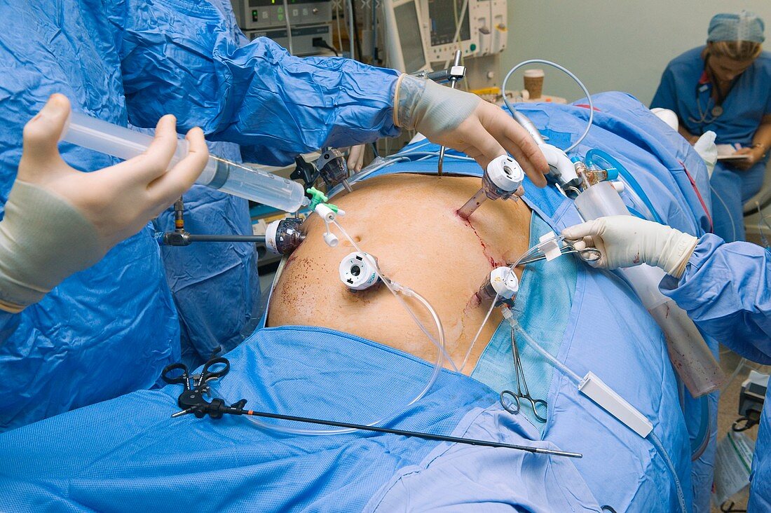
| MODEL RELEASED. Oesophageal cancer surgery. Image 8 of 28. Surgeon using a syringe to flush out the intestines while using laparoscopic tools inside the abdomen of a patient being operated on for cancer of the oesophagus (gullet). This flushing dilates the intestines,making it easier to advance the instruments and tubes being used. The operation was carried out in two stages. First was laparoscopic keyhole surgery inside the abdomen (seen here),to operate on the lower,abdominal part of the cancer. This was followed by open-chest surgery (thoracotomy) to remove the cancerous oesophageal tissue and connect the remaining healthy part back to the stomach (oesophago-gastric anastomosis) | |
| Lizenzart: | Lizenzpflichtig |
| Credit: | Science Photo Library / Marazzi, Dr. P. |
| Bildgröße: | 5147 px × 3425 px |
| Modell-Rechte: | vorhanden |
| Eigentums-Rechte: | nicht erforderlich |
| Restrictions: |
|
Preise für dieses Bild ab 15 €
Universitäten & Organisationen
(Informationsmaterial Digital, Informationsmaterial Print, Lehrmaterial Digital etc.)
ab 15 €
Redaktionell
(Bücher, Bücher: Sach- und Fachliteratur, Digitale Medien (redaktionell) etc.)
ab 30 €
Werbung
(Anzeigen, Aussenwerbung, Digitale Medien, Fernsehwerbung, Karten, Werbemittel, Zeitschriften etc.)
ab 55 €
Handelsprodukte
(bedruckte Textilie, Kalender, Postkarte, Grußkarte, Verpackung etc.)
ab 75 €
Pauschalpreise
Rechtepakete für die unbeschränkte Bildnutzung in Print oder Online
ab 495 €
Keywords
- Abdomen,
- Arthroskopie,
- Arzt,
- Ärzte,
- Ausrüstung,
- Bauch,
- Behandlung,
- behandschuht,
- Betrieb,
- Chirurg,
- Chirurgen,
- chirurgisch,
- Close-up,
- Darm-,
- Detail,
- einspritzend,
- Endoskop,
- Endoskopie,
- endoskopisch,
- Erwachsene,
- Fluid,
- Flüssigkeit,
- Gedärme,
- geduldig,
- Gerät,
- Hand,
- Hände,
- Handschuh,
- Handschuhe,
- Instrumente,
- kaukasisch,
- Krankenhaus,
- Laparoskop,
- Laparoskopie,
- Medizin,
- medizinisch,
- Mensch,
- Menschen,
- minimal-invasive,
- Oesophagus,
- Operation,
- Operationssaal,
- Person,
- schützend,
- Speiseröhre,
- Spritze,
- steril,
- Technologie,
- technologisch,
- Wasser,
- weiß,
- Werkzeuge