Ischaemia,angiogram
Bildnummer 11553193
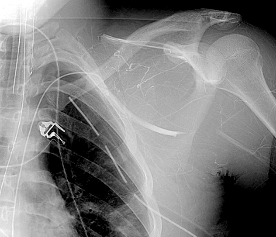
| Ischaemia,angiogram. Front view of ischaemia (lack of blood flow) in the left shoulder of a 21 year old patient. There are fewer blood vessels visible compared to normal,which is due to a blocked left axillary artery (thicker white line that ends abruptly at centre right) resulting from a road traffic accident. The blockage may be due to a thrombus (blood clot) or embolism (air bubble or loose blood clot). A chest drain (bottom right to upper centre left) and an intubation catheter (vertical lines at top left) are also seen. At centre left is an ECG electrode. An angiogram is an X-ray technique that uses a contrast dye to highlight blood vessels | |
| Lizenzart: | Lizenzpflichtig |
| Credit: | Science Photo Library / Zephyr |
| Bildgröße: | 4550 px × 3912 px |
| Modell-Rechte: | nicht erforderlich |
| Eigentums-Rechte: | nicht erforderlich |
| Restrictions: | - |
Preise für dieses Bild ab 15 €
Universitäten & Organisationen
(Informationsmaterial Digital, Informationsmaterial Print, Lehrmaterial Digital etc.)
ab 15 €
Redaktionell
(Bücher, Bücher: Sach- und Fachliteratur, Digitale Medien (redaktionell) etc.)
ab 30 €
Werbung
(Anzeigen, Aussenwerbung, Digitale Medien, Fernsehwerbung, Karten, Werbemittel, Zeitschriften etc.)
ab 55 €
Handelsprodukte
(bedruckte Textilie, Kalender, Postkarte, Grußkarte, Verpackung etc.)
ab 75 €
Pauschalpreise
Rechtepakete für die unbeschränkte Bildnutzung in Print oder Online
ab 495 €
Keywords
- 20er Jahre,
- abnormal,
- Angiografie,
- Angiogramm,
- Arterie,
- arteriell,
- Beschränkung,
- Bildgebung,
- Blockierung,
- Blutfluss,
- Blutgefäß,
- Blutversorgung,
- CVA,
- Diagnose,
- digitale Angiographie,
- Einfarbig,
- Erwachsene,
- Gesundheitswesen,
- intubiert,
- Ischämie,
- Kondition,
- Medizin,
- medizinisch,
- Mensch,
- menschlicher Körper,
- Obstruktion,
- Person,
- Radiographie,
- Röntgen,
- Röntgengerät,
- rta,
- Schlaganfall,
- Schulter,
- Schwarz und weiß,
- Störung,
- Thorax,
- Trauma,
- traumatisch,
- Truhe,
- Tube,
- ungesund,
- vaskulär,
- verletzt,
- Verletzung,
- verstopft,
- Vorderansicht,
- zwanziger Jahre