Small intestine lining,light micrograph
Bildnummer 11550593
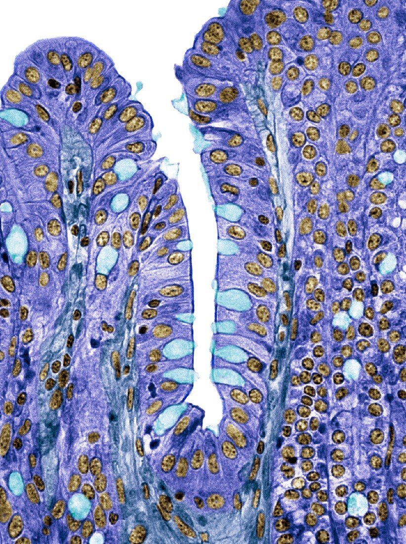
| Small intestine lining. Light micrograph of a section through the finger-like projections (villi) of the duodenum,the uppermost part of the small intestine. These increase the surface area for the absorption of food. Within the columnar epithelium of the outer surface (purple) are goblet cells (light blue),which secrete mucus to lubricate food and prevent self-digestion. The lamina propria (central core,dark blue) contains the blood supply that transports the products of digestion. Magnification: x350 when printed at 10 centimetres wide | |
| Lizenzart: | Lizenzpflichtig |
| Credit: | Science Photo Library / Gschmeissner, Steve |
| Bildgröße: | 3662 px × 4900 px |
| Modell-Rechte: | nicht erforderlich |
| Eigentums-Rechte: | nicht erforderlich |
| Restrictions: | - |
Preise für dieses Bild ab 15 €
Universitäten & Organisationen
(Informationsmaterial Digital, Informationsmaterial Print, Lehrmaterial Digital etc.)
ab 15 €
Redaktionell
(Bücher, Bücher: Sach- und Fachliteratur, Digitale Medien (redaktionell) etc.)
ab 30 €
Werbung
(Anzeigen, Aussenwerbung, Digitale Medien, Fernsehwerbung, Karten, Werbemittel, Zeitschriften etc.)
ab 55 €
Handelsprodukte
(bedruckte Textilie, Kalender, Postkarte, Grußkarte, Verpackung etc.)
ab 75 €
Pauschalpreise
Rechtepakete für die unbeschränkte Bildnutzung in Print oder Online
ab 495 €
Keywords
- absondern,
- Absorption,
- Anatomie,
- anatomisch,
- Bindegewebe,
- Biologie,
- biologisch,
- Darm,
- Darm-,
- Dünndarm,
- eingefärbt,
- Epithel,
- epithelial,
- farbig,
- Futter,
- gastrointestinal,
- gefärbt,
- gesund,
- GI tract,
- Histologie,
- histologisch,
- Ileum,
- Jejunum,
- Kelch,
- Lichtmikroskop,
- lichtmikroskopische Aufnahme,
- Magen-Darm,
- Membran,
- menschlicher Körper,
- Mikrovilli,
- Mikrovillus,
- normal,
- Person,
- Querschnitt,
- Säule,
- Schleimhaut,
- sekretorisch,
- Sektion,
- sektioniert,
- Struktur,
- Verdauung,
- Verdauungskanal,
- Verdauungstrakt,
- Zelle,
- Zellen,
- Zotte,
- Zotten