Chicken embryo,light micrograph
Bildnummer 11549559
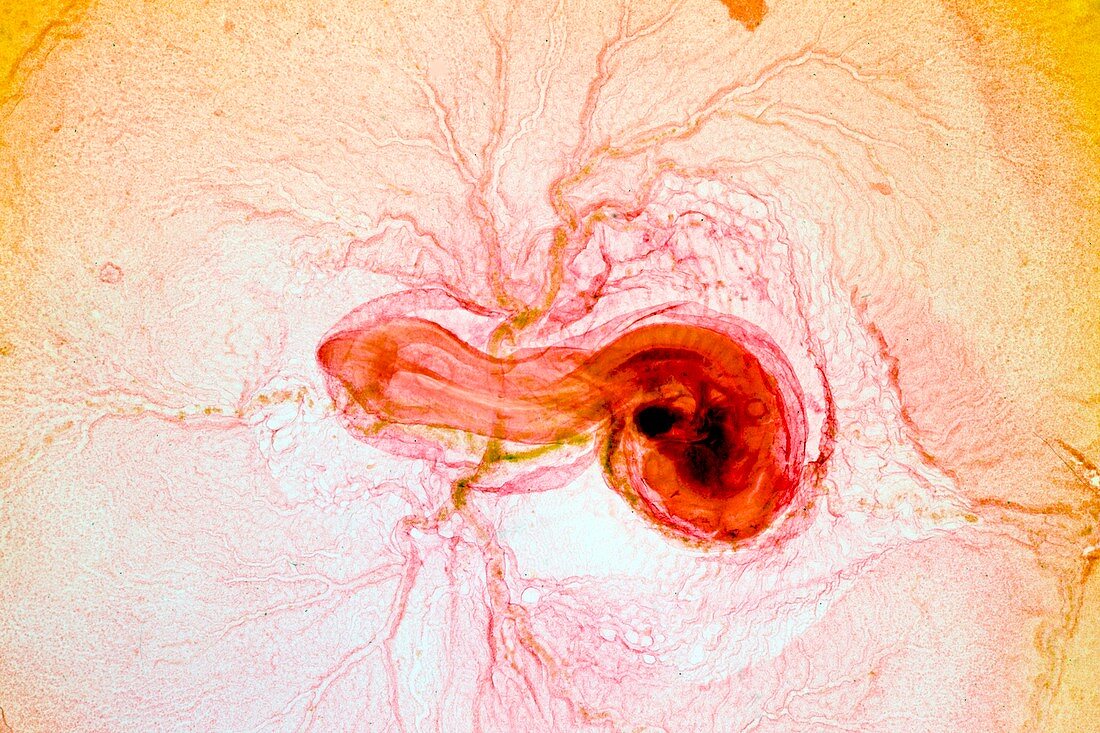
| Chicken embryo. Light micrograph of the embryo of a chicken (Gallus domestica) after 3 days of development,surrounded by the amniotic membrane and the yolk blood vessels. The head area (right) includes the swollen anterior bulbous forebrain (cerebral hemisphere),a fully developed eye with lens (black,centre of head). The heart (also black) is to the left of the eye. The brain continues as the mid and hind-brain and then into the body as the neural tube (with 35 somites,muscle blocks,along either side) ending in the tail (left). Magnification: x14 when printed at 10 centimetres across | |
| Lizenzart: | Lizenzpflichtig |
| Credit: | Science Photo Library / Wheeler, Dr. Keith |
| Bildgröße: | 5616 px × 3744 px |
| Modell-Rechte: | nicht erforderlich |
| Eigentums-Rechte: | nicht erforderlich |
| Restrictions: | - |
Preise für dieses Bild ab 15 €
Universitäten & Organisationen
(Informationsmaterial Digital, Informationsmaterial Print, Lehrmaterial Digital etc.)
ab 15 €
Redaktionell
(Bücher, Bücher: Sach- und Fachliteratur, Digitale Medien (redaktionell) etc.)
ab 30 €
Werbung
(Anzeigen, Aussenwerbung, Digitale Medien, Fernsehwerbung, Karten, Werbemittel, Zeitschriften etc.)
ab 55 €
Handelsprodukte
(bedruckte Textilie, Kalender, Postkarte, Grußkarte, Verpackung etc.)
ab 75 €
Pauschalpreise
Rechtepakete für die unbeschränkte Bildnutzung in Print oder Online
ab 495 €
Keywords
- Auge,
- Biologie,
- biologisch,
- Blutgefäß,
- Drei,
- Eigelb,
- einer,
- Embryo,
- Embryologie,
- Entwicklung,
- Entwicklungsbiologie,
- Fauna,
- Fluid,
- Gefäße,
- Gehirn,
- Hähnchen,
- Herz,
- Kopf,
- Lichtmikroskop,
- lichtmikroskopische Aufnahme,
- Linse,
- Natur,
- Nervensystem,
- neural,
- Organ,
- Organe,
- ornithologisch,
- Reproduktion,
- reproduktiv,
- Single,
- Somiten,
- Stunde,
- Tier,
- Tierwelt,
- Vene,
- Venen,
- Vogel,
- Vogelkunde,
- zentrales Nervensystem,
- Zoologie,
- zoologisch