Dogfish gill,light micrograph
Bildnummer 11549533
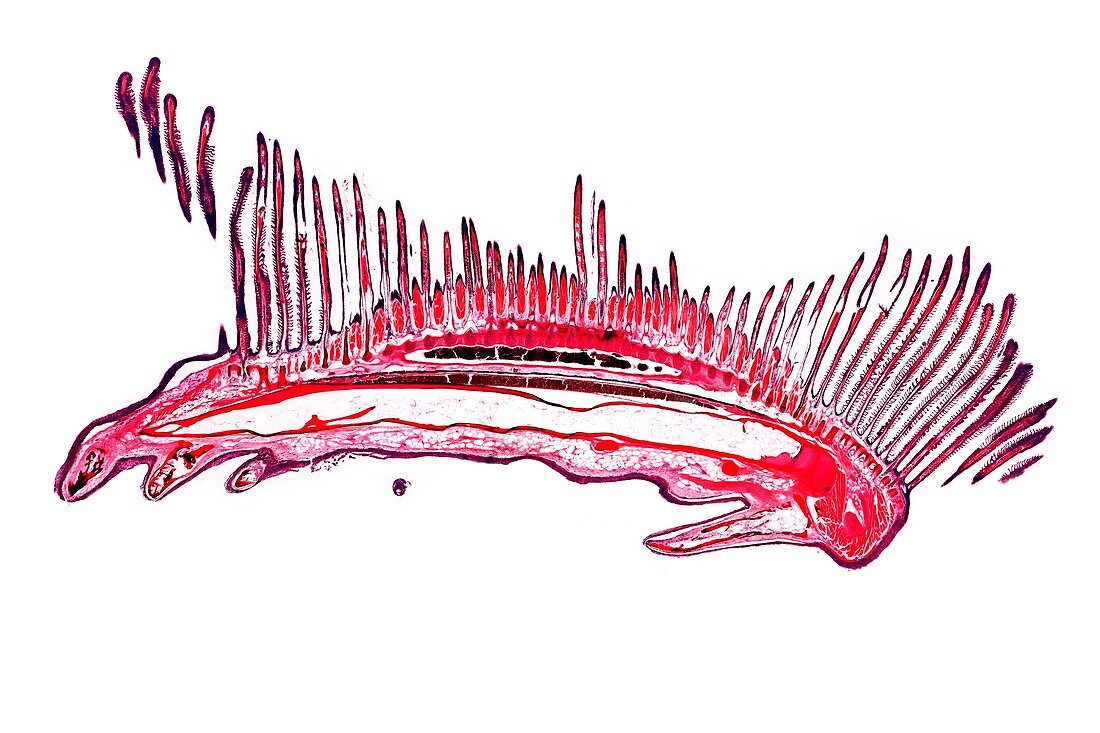
| Dogfish gill. Light micrograph of a gill ray and gill lamellae,from a dogfish (Scyliorhinus canicular). This section through the gill shows parts of its cartilaginous support structure. Across top is the gill arch and the gill rays,while the pharyngeal wall is across centre. Colours of tissues are: epidermis (purple),muscles (red),blood cells (purple-black). Cut lengthwise across bottom are the afferent and efferent brachial arteries taking blood to and from the gills. Gills are used by fish to breathe underwater,to extract oxygen from the water,and to dispose of waste carbon dioxide. Magnification: x36 when printed at 10 centimetres across | |
| Lizenzart: | Lizenzpflichtig |
| Credit: | Science Photo Library / Wheeler, Dr. Keith |
| Bildgröße: | 5616 px × 3744 px |
| Modell-Rechte: | nicht erforderlich |
| Eigentums-Rechte: | nicht erforderlich |
| Restrictions: | - |
Preise für dieses Bild ab 15 €
Universitäten & Organisationen
(Informationsmaterial Digital, Informationsmaterial Print, Lehrmaterial Digital etc.)
ab 15 €
Redaktionell
(Bücher, Bücher: Sach- und Fachliteratur, Digitale Medien (redaktionell) etc.)
ab 30 €
Werbung
(Anzeigen, Aussenwerbung, Digitale Medien, Fernsehwerbung, Karten, Werbemittel, Zeitschriften etc.)
ab 55 €
Handelsprodukte
(bedruckte Textilie, Kalender, Postkarte, Grußkarte, Verpackung etc.)
ab 75 €
Pauschalpreise
Rechtepakete für die unbeschränkte Bildnutzung in Print oder Online
ab 495 €
Keywords
- Anatomie,
- anatomisch,
- Arterie,
- atmen,
- Atmung,
- Atmungssystem,
- ausgeschnitten,
- Ausschnitte,
- Biologie,
- biologisch,
- Blut,
- Blutgefäß,
- Blutzellen,
- Epidermis,
- Fauna,
- Fisch,
- Fischkunde,
- Gefäße,
- Kiemen,
- Kohlendioxid,
- Lichtmikroskop,
- lichtmikroskopische Aufnahme,
- Meeresbiologie,
- Muskeln,
- Natur,
- Sektion,
- sektioniert,
- Strahlen,
- Tier,
- Tierwelt,
- vaskulär,
- Zoologie,
- zoologisch