Developing heart muscle cell
Bildnummer 11549213
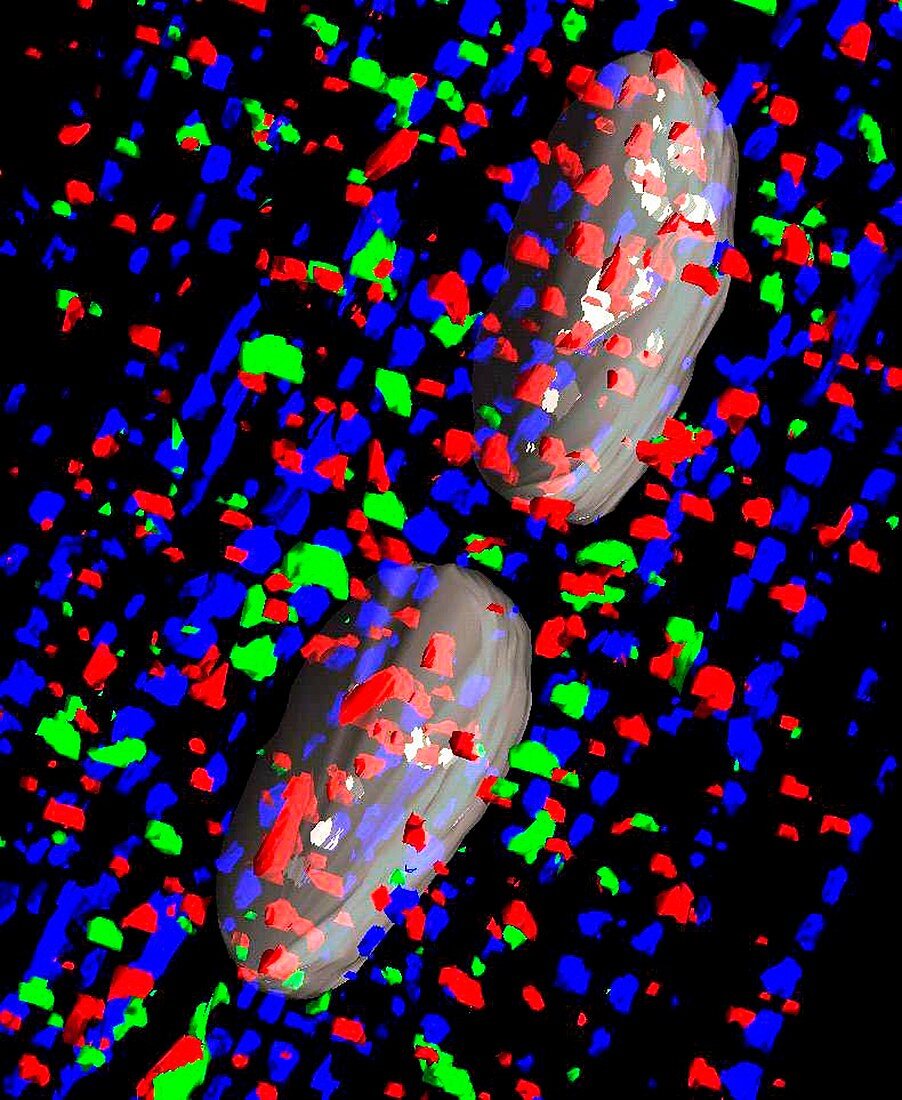
| Developing heart muscle cell. 3D composite image showing the two nuclei (large white) and synthesised proteins of a developing binucleate cardiomyocyte. The image is made up of multiple fluorescence deconvolution micrograph images. Myosin (blue) will be used to form contractile fibres (myofibrils),and the receptor proteins (red and green) will go on to form chemical receptors within the cell. Magnification: x600 when printed at 10 centimetres wide | |
| Lizenzart: | Lizenzpflichtig |
| Credit: | Science Photo Library / R. BICK, B. POINDEXTER, UT MEDICAL SCHOOL |
| Bildgröße: | 3798 px × 4630 px |
| Modell-Rechte: | nicht erforderlich |
| Eigentums-Rechte: | nicht erforderlich |
| Restrictions: | - |
Preise für dieses Bild ab 15 €
Universitäten & Organisationen
(Informationsmaterial Digital, Informationsmaterial Print, Lehrmaterial Digital etc.)
ab 15 €
Redaktionell
(Bücher, Bücher: Sach- und Fachliteratur, Digitale Medien (redaktionell) etc.)
ab 30 €
Werbung
(Anzeigen, Aussenwerbung, Digitale Medien, Fernsehwerbung, Karten, Werbemittel, Zeitschriften etc.)
ab 55 €
Handelsprodukte
(bedruckte Textilie, Kalender, Postkarte, Grußkarte, Verpackung etc.)
ab 75 €
Pauschalpreise
Rechtepakete für die unbeschränkte Bildnutzung in Print oder Online
ab 495 €
Keywords
- 3-d,
- 3-dimensional,
- 3D,
- Anatomie,
- Atomkern,
- befleckt,
- Biologie,
- biologisch,
- Dreidimensional,
- Eiweiß,
- Entwicklung,
- Fluoreszenzentfaltung,
- gesund,
- Herz,
- Histologie,
- histologisch,
- Jung,
- Kerne,
- konfokal,
- Lichtmikroskop,
- lichtmikroskopische Aufnahme,
- Mensch,
- Mikroskopie,
- Muskel,
- Muskeln,
- normal,
- Person,
- Proteine,
- Verfärbung,
- Zellbilogie,
- Zusammengesetzt,
- Zytologie