Small intestine,fluorescence micrograph
Bildnummer 11549211
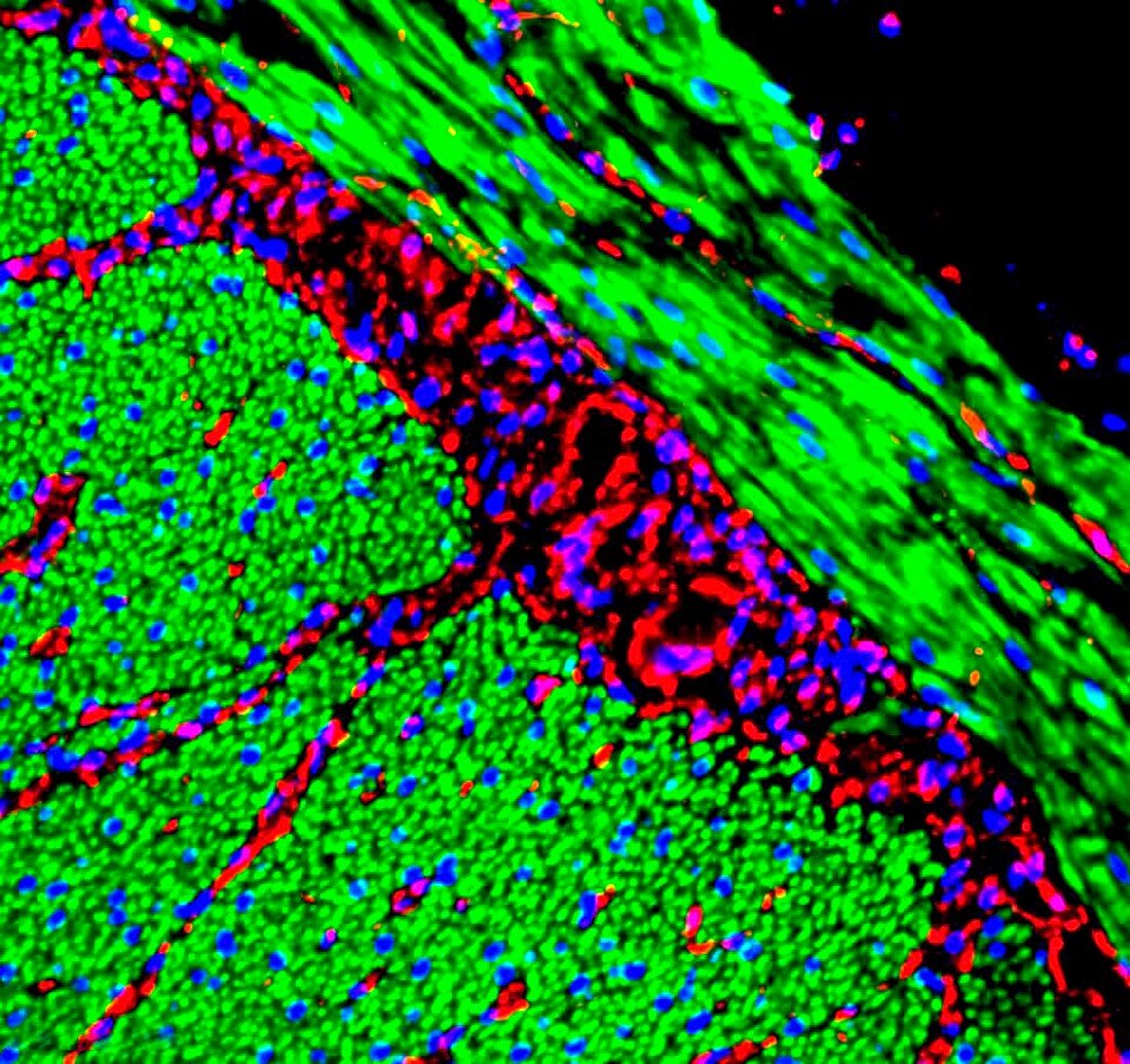
| Small intestine. Fluorescence deconvolution micrograph showing a section through the myenteric,or Auerbach's,plexus of the small intestine. Smooth muscle cell nuclei,in the muscularis externa (diagonal,top right),and axonal cell nuclei are shown in blue. Red shows calcium-sensing proteins. This network of cells controls local intestinal wall movement. When the wall is stretched by its contents,nerves in the plexus cause the muscles (green) above the dilation to contract and those below it to relax. This results in a wave-like movement of the gut wall (peristalsis) which squeezes the contents along the intestine. Magnification: x400 when printed at 10 centimetres wide | |
| Lizenzart: | Lizenzpflichtig |
| Credit: | Science Photo Library / R. BICK, B. POINDEXTER, G. KLEIN (UTMB GALVESTON), UT MEDICAL SCHOOL |
| Bildgröße: | 4319 px × 4063 px |
| Modell-Rechte: | nicht erforderlich |
| Eigentums-Rechte: | nicht erforderlich |
| Restrictions: | - |
Preise für dieses Bild ab 15 €
Universitäten & Organisationen
(Informationsmaterial Digital, Informationsmaterial Print, Lehrmaterial Digital etc.)
ab 15 €
Redaktionell
(Bücher, Bücher: Sach- und Fachliteratur, Digitale Medien (redaktionell) etc.)
ab 30 €
Werbung
(Anzeigen, Aussenwerbung, Digitale Medien, Fernsehwerbung, Karten, Werbemittel, Zeitschriften etc.)
ab 55 €
Handelsprodukte
(bedruckte Textilie, Kalender, Postkarte, Grußkarte, Verpackung etc.)
ab 75 €
Pauschalpreise
Rechtepakete für die unbeschränkte Bildnutzung in Print oder Online
ab 495 €
Keywords
- Anatomie,
- Atomkern,
- Axon,
- axonal,
- Axone,
- befleckt,
- Biologie,
- biologisch,
- Darm,
- Darm-,
- Dünndarm,
- Eingeweide,
- Eiweiß,
- Fasern,
- Fluoreszenzentfaltung,
- Gastroenterologie,
- gesund,
- Histologie,
- histologisch,
- Kerne,
- konfokal,
- Kontrolle,
- Lichtmikroskop,
- lichtmikroskopische Aufnahme,
- Mensch,
- Mikroskopie,
- Motilität,
- Netzwerk,
- normal,
- Person,
- Plexus myentericus,
- Sektion,
- sektioniert,
- Verdauungssystem,
- Verfärbung,
- Zellbilogie,
- Zelle,
- Zellen,
- Zytologie